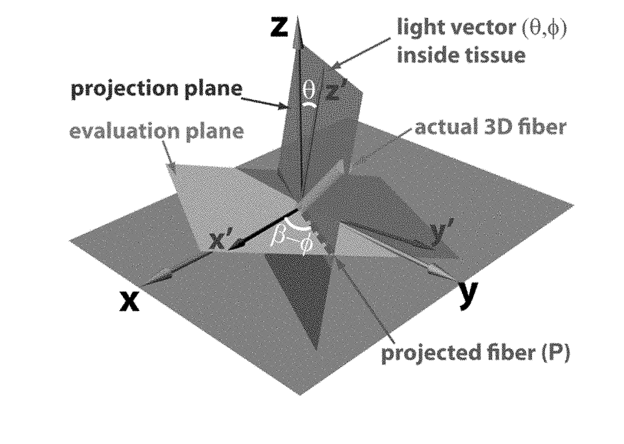
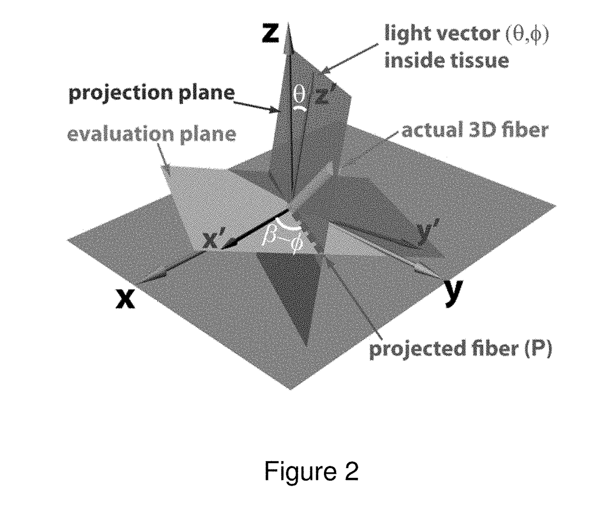
Optical polarization tractography methods
a fiber structure and optical polarization technology, applied in the field of optical polarization tractography methods and systems, can solve the problems of fibrous structure changes under diseased and pathological conditions, dti suffers from a spatial resolution largely limited to the submillimeter range, time-consuming, laborious, etc., and achieves high imaging speed, large imaging area, and unprecedented details at the cellular resolution level.
- Summary
- Abstract
- Description
- Claims
- Application Information
AI Technical Summary
Benefits of technology
Problems solved by technology
Method used
Image

Examples
example 1
Demonstration of the 3D OPT System in a Small Piece of the Mouse EDL Muscle
[0073]The dual-angle 3D OPT system and method according to an embodiment of the invention was demonstrated by imaging a small piece of the mouse EDL muscle which was placed at known orientations in the space. The middle of the EDL muscle had relatively homogeneous myofiber orientation. The muscle sample was fixed in 10% paraformaldehyde after excising it from the animal.
[0074]FIG. 4(a) illustrates the geometry of the EDL muscle in the dual-angle OPT imaging wherein the EDL sample was rotated in the laboratory coordinates by γ1 and γ2 around the x-axis to complete the dual angle imaging. The muscle sample was mounted on a two-axis rotation stage carefully aligned with the scanning directions to provide accurate rotation around the x-axis (C-scan) and y-axis (B-scan). To implement the dual-angle scanning, the EDL muscle was first scanned at γ1=−20° respect to the x-axis (C-scan); then imaged again at γ2=+20° af...
example 2
Imaging 3D Fiber Orientation in the Mouse TA Muscle
[0085]In order to further evaluate the accuracy of the 3D measurements according to an embodiment of the OPT system of the invention, a piece of fixed mouse TA muscle was imaged. FIG. 7(a) shows the 3D intensity images of the TA muscle imaged at γ2=+25°, where fibers are difficult to observe. In sharp contrast, the corresponding 3D tractography (FIG. 7(b)) constructed in dual-angle OPT clearly revealed 3D fiber structure. The 3D tractography indicated that the muscle fibers were mostly aligned along the long axis of the TA muscle.
[0086]FIG. 7(c) demonstrates the capability of 3D OPT in revealing detailed fiber architecture by selectively revealing two fiber bundles at two separate locations inside the muscle. As expected, the two muscle bundles were clearly organized along the long axis of the muscle toward the apex. FIG. 7(d) further demonstrated that the details of the fiber bundles can be studied in detail by projecting the 3D fi...
example 3
Imaging 3D Fiber Orientation in Cartilage
[0087]In this example, dual-angle OPT was applied to image the collagen fiber organization in a piece of bovine articular cartilage. The cartilage sample was excised from the middle phalanx and fixed in 4% paraformaldehyde. This sample was imaged from the cartilage surface (referred to as the “top-scan”) using the dual-angle procedures at γ1=−25° first and then at γ2=+25°.
[0088]No fibers can be observed from the 3D intensity image of the cartilage sample (FIG. 8(a)). However, the constructed 3D fiber tractography from dual-angle OPT (FIG. 8(b)) clearly revealed the unique arcade-like fiber organization in the cartilage. In the “superficial” zone, fibers were oriented relatively parallel to the sample surface. Then the fibers arched in the “transitional” zone to be eventually perpendicular toward the boundary between non-calcified and calcified cartilage in the deeper “radial” zones. Moreover, the 3D tractography correctly revealed the “brushi...
PUM
| Property | Measurement | Unit |
|---|---|---|
| central wavelength | aaaaa | aaaaa |
| central wavelength | aaaaa | aaaaa |
| central wavelength | aaaaa | aaaaa |
Abstract
Description
Claims
Application Information
 - R&D
- Intellectual Property
- Life Sciences
- Materials
- Tech Scout
- Unparalleled Data Quality
- Higher Quality Content
- 60% Fewer Hallucinations
Browse by: Latest US Patents, China's latest patents, Technical Efficacy Thesaurus, Application Domain, Technology Topic, Popular Technical Reports.
© 2025 PatSnap. All rights reserved.Legal|Privacy policy|Modern Slavery Act Transparency Statement|Sitemap|About US| Contact US: help@patsnap.com