Cytotoxicity mediation of cells evidencing surface expression of MCSP
A cytotoxic and cell-based technology, applied in the field of diagnosis and treatment of cancerous diseases, can solve the problems of undisclosed monoclonal antibodies
- Summary
- Abstract
- Description
- Claims
- Application Information
AI Technical Summary
Problems solved by technology
Method used
Image
Examples
Embodiment 1
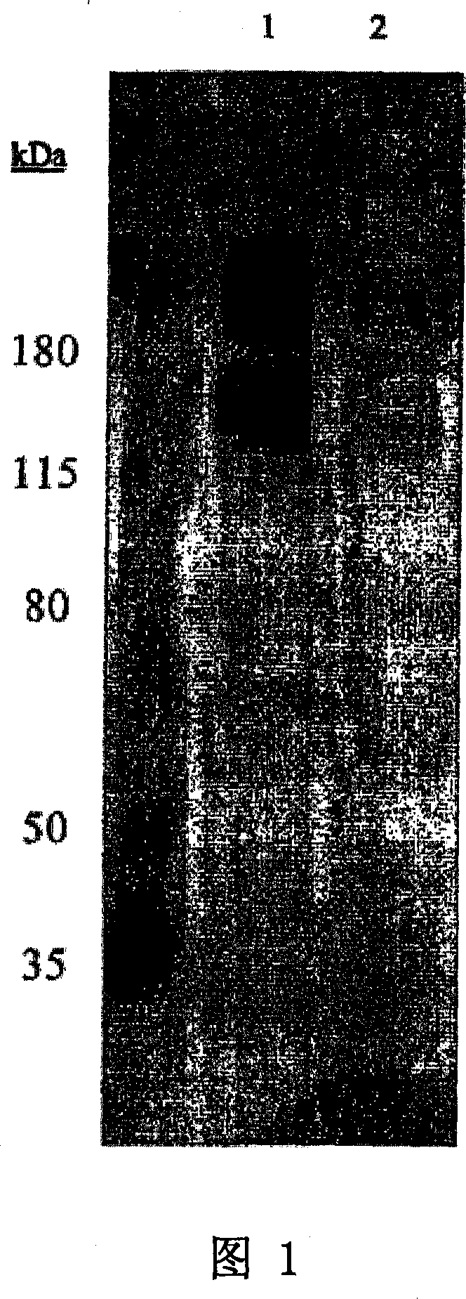
[0075] Identification of binding proteins by Western blotting
[0076] To identify the antigen bound by antibody 11BD-2E11-2, cell membranes expressing the antigen were subjected to gel electrophoresis and transferred to the membrane by Western blotting to determine the proteins detected by the antibody.
[0077] 1: Membrane Preparation
[0078] Previous work has shown that 11BD-2E11-2 binds to the breast cancer cell line MDA-MB-231 (MB-231) by flow cytometry. Previous work also showed the effectiveness of 11BD-2E11-2 against the ovarian cancer cell line OVCAR-3. Therefore, antigen identification was performed using membrane preparations of these two cell lines. Whole-cell membranes were prepared from confluent cultures of MB-231 breast cancer or OVCAR-3 ovarian cells. The medium was removed from the cell stack and the cells were washed with phosphate buffered saline. Cells were dissociated with dissociation buffer (Gibco-BRL, Grand Island, NY) for 20 minutes at 37°C on a ...
Embodiment 2
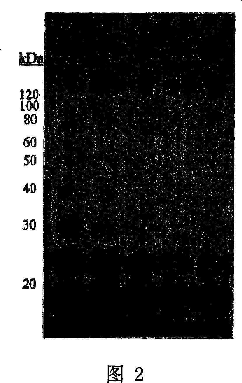
[0082] Determination of glycosylation of antigen bound by 11BD-2E11-2
[0083] To determine whether the antigen recognized by the 11BD-2E11-2 antibody was a glycoprotein, MB-231 membrane protein was treated with different combinations of PNGase F, endo-o-glycosidase, sialidase, galactosidase and glucosamine Glycosidase co-incubation. Membranes were separated by SDS-PAGE and then Western blotted as done with 11BD-2E11-2. Figure 2 shows the results of 11BD-2E11-2 binding to MDA-MB-231 membranes in: a separate deglycosylation buffer (lane 1), with PNGase F, endo- o-glucosidase, sialidase, galactosidase and glucosaminidase (glucosaminidase) in combination (lane 2), and PNG enzyme, endo-o-glucosidase and sialidase in combination (lane 3 ), sialidase alone (lane 4), endo-o-glycosidase alone (lane 5), and PNGase alone (lane 6). Treatment of MB-231 membrane with glycosidase did not eliminate the binding of 11BD-2E11-2, but a shift in protein molecular weight was found in all lanes,...
Embodiment 3
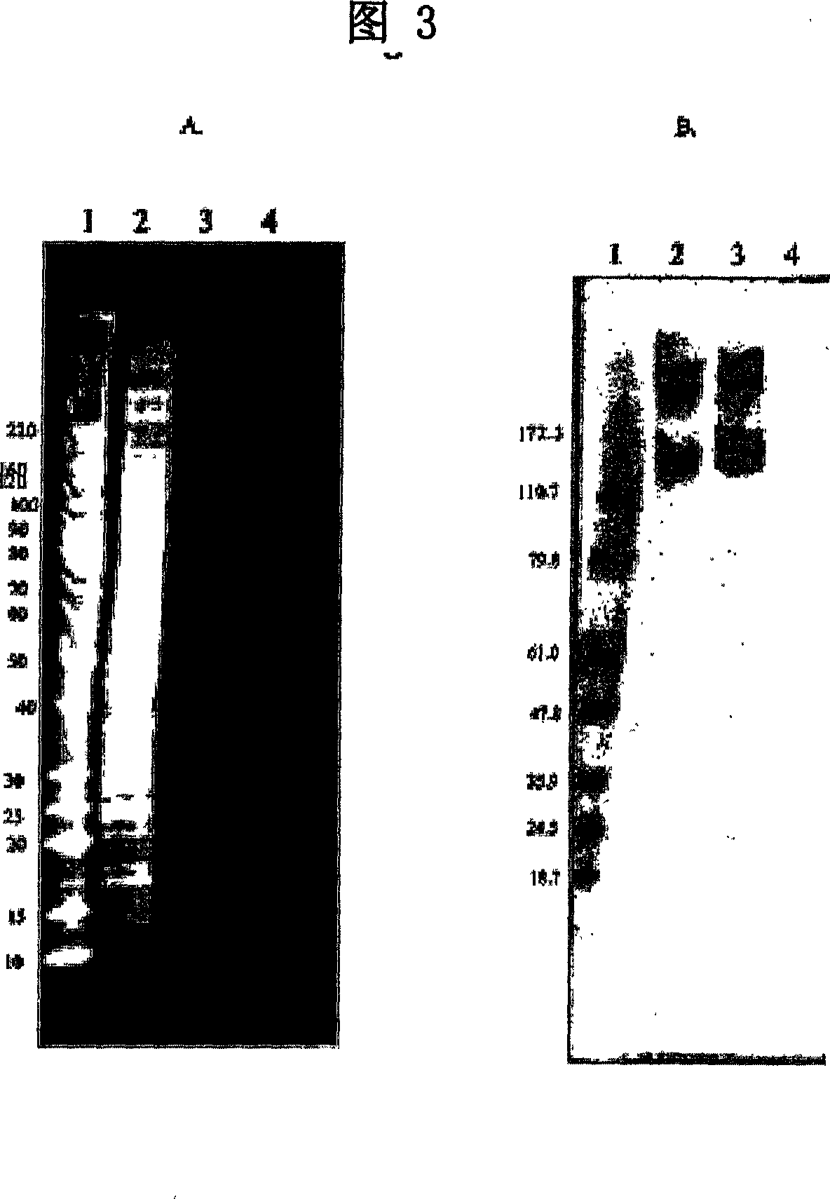
[0085] Identification of 11BD-2E11-2 binding antigen
[0086] 1. Immunoprecipitation
[0087] Cognate ligands were isolated by immunoprecipitation of solubilized membrane glycoproteins with 11BD-2E11-2 antibody to identify the antigen corresponding to 11BD-2E11-2. 100 μL of Protein G Dynabeads (Dynal Biotech, Lake Success NY) were washed 3 times with 1 mL of 0.1 M sodium phosphate buffer, pH 6.0. 100 μg of 11BD-2E11-2 dissolved in pH 6.0 0.1 M sodium phosphate buffer in a total volume of 100 μL was added to the washed beads. The mixture was incubated for 1 hour with rotational mixing. Unbound antibody was removed, and the 11BD-2E11-2-coated antibody was washed three times with 0.5 mL of a pH 7.4 sodium phosphate solution containing 0.1% Tween-20. The 11BD-2E11-2-coated antibody was washed twice with 1 mL of 0.2 M pH 8.2 triethanolamine. 1 mL of 0.02 M dimethylpimelimidate in 0.2 M triethanolamine (pH 8.2) was added and incubated for 30 minutes with swirling mixing to chemi...
PUM
 Abstract
Description
Claims
Application Information
 - R&D
- Intellectual Property
- Life Sciences
- Materials
- Tech Scout
- Unparalleled Data Quality
- Higher Quality Content
- 60% Fewer Hallucinations
Browse by: Latest US Patents, China's latest patents, Technical Efficacy Thesaurus, Application Domain, Technology Topic, Popular Technical Reports.
© 2025 PatSnap. All rights reserved.Legal|Privacy policy|Modern Slavery Act Transparency Statement|Sitemap|About US| Contact US: help@patsnap.com


