
Method for preparing epithelium of autologous cornea

A corneal epithelial and autologous technology, applied in biochemical equipment and methods, artificial cell constructs, microorganisms, etc., can solve the problems of slow degradation, increased patient pain, and surgical failure, achieving good results and flexible operations

Active Publication Date: 2007-02-21
北京智慧细胞生物科技有限公司
View PDF1 Cites 76 Cited by
  • Summary
  • Abstract
  • Description
  • Claims
  • Application Information

AI Technical Summary

Problems solved by technology

[0004] However, there are many deficiencies in the bioengineered corneal epithelium of the embryonic limbal tissue: first, because the embryonic limbal tissue is derived from a foreign body, the corneal epithelium prepared from it will have immune rejection with the immune cells in the patient's limbus, thereby increasing the Patient distress and likelihood of surgical failure
Second, because the bio-scaffolds used to prepare the cornea are collagen, contact lenses or amnion, it is difficult for corneal stem cells and epithelial cells to grow uniformly on the surface, and the degradation rate after surgery is slow, thus limiting clinical application
The tissue-engineered cornea overcomes the above shortcomings, but when there are problems in both corneas, the source of autologous limbal stem cells is missing

Method used

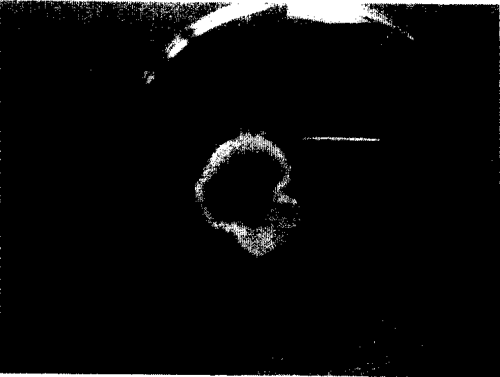
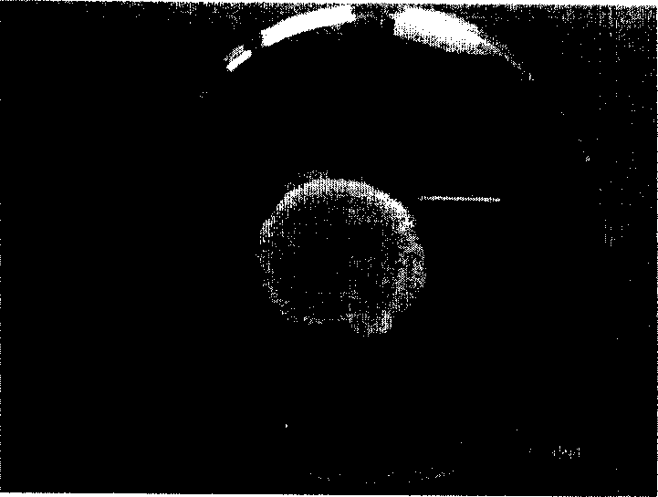
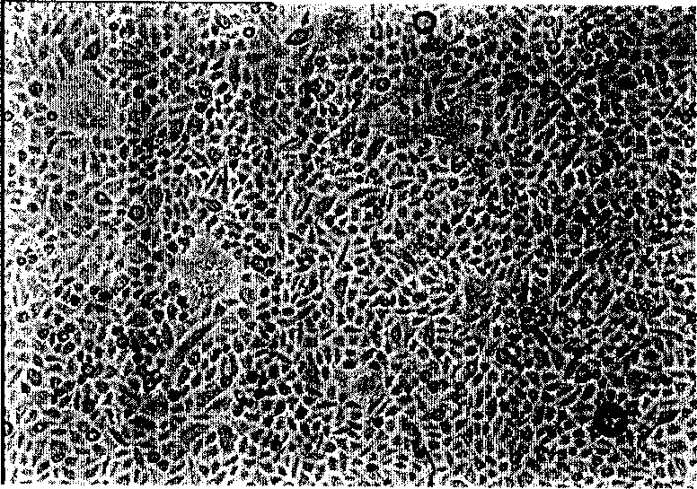

Image

  • Method for preparing epithelium of autologous cornea
  • Method for preparing epithelium of autologous cornea
  • Method for preparing epithelium of autologous cornea

Examples


Embodiment 1

[0034] Example 1: Preparation of tissue-engineered autologous corneal epithelium derived from oral mucosa

[0035] 1. Extraction and primary culture of healthy cells from oral mucosa tissue

[0036] 1. Material collection in the operating room (100-level air purification): Professional doctors take tissue pieces of 1mm×3mm from the patient’s sublingual oral mucosa.

[0037] 2. Preservation of the taken tissue: During transportation to the laboratory, the tissue block can be stored (4° C.) in a 15 ml preservation tube containing a preservation solution containing DMEM and 10% serum.

[0038] 3. Sterilize, clean, and trim tissue in an ultra-clean workbench: take fresh tissue or preserved tissue, wash it with PBS (phosphate buffered saline) containing 100mg / ml gentamicin for 3 times, and trim excess fat and subcutaneous tissue connective tissue.

[0039] 4. Tissue digestion (Thermo CO 2 Incubator): Place the above tissue block in a 35mm petri dish, add trypsin / EDTA solution wi...

Embodiment 2

[0060] Example 2: Preparation of limbal cell-derived tissue-engineered autologous corneal epithelium

[0061] 1. Extraction and primary culture of healthy cells from limbal tissue

[0062] 1. Material collection in the operating room (100-level air purification): Professional doctors take tissue pieces of 1mm×1mm size from the limbus of the normal eye on the other side of the patient.

[0063] 2. Preservation of the taken tissue: During transportation to the laboratory, the tissue block can be stored (4° C.) in a 15 ml preservation tube containing a preservation solution containing DMEM and 10% serum.

[0064] 3. Sterilize, clean, and trim tissue in an ultra-clean workbench: take fresh tissue or preserved tissue, wash it with PBS containing 100mg / ml gentamicin for 3 times, and trim excess fat and subcutaneous connective tissue.

[0065] 4. Tissue digestion (Thermo CO 2 Incubator): Place the above tissue block in a 35mm petri dish, add trypsin / EDTA solution with a concentrati...


PUM


Abstract

This invention discloses a method for preparing auto-corneal epithelia. The method comprises: (1) performing primary culture of cells of patients' oral mucosa tissue or corneal limbal tissue for 3-15 days; (2) performing proliferation sub-culture for 7-30 days; (3) using fibrinogen as the supporting material to construct tissue engineering corneal epithelia together with the proliferation sub-cultured cells. The method can avoid the problems of immunological rejection, pollution, ununiform cell growth caused by the biological support, and slow degradation faced by presents techniques. The preparation of auto-corneal in this invention is not restricted by corneal epithelia damage, and suitable corneal is available for patients at any situation.

Description

technical field [0001] The invention relates to a preparation method of autologous corneal epithelium. Background technique [0002] Burns, chemical burns, ocular surface tumors, allergic or radiation injuries, hereditary eye diseases, ocular surface suppuration, and other eye syndromes can cause corneal epithelial cells to shed, and make the conjunctiva cover the corneal surface, resulting in a large number of new blood vessels in the cornea Corneal diseases such as hyperplasia, recurrent pterygium and even blindness. For the treatment of such ophthalmic diseases, surgical excision is generally required, but recurrence and hyperplasia still occur after excision. Therefore, to treat such diseases fundamentally, it is necessary to construct the tissue structure of the cornea and supplement the stem cells of the limbus. [0003] In order to solve such problems, people have invented a bioengineered cornea, which is constructed on a biological scaffold by culturing corneal ste...

Claims


Application Information

Patent Timeline
IPC IPC(8): C12N5/08C12N5/071
Inventor 韩斌
Owner 北京智慧细胞生物科技有限公司
Learn More