
Protein modified microsphere and application thereof

A protein modification and microsphere technology, applied in the field of biomedical engineering, can solve the problems of over-differentiated stem cells, poor reproducibility, and cumbersome culture operations.

Active Publication Date: 2022-08-05
TSINGHUA UNIV
View PDF0 Cites 0 Cited by
  • Summary
  • Abstract
  • Description
  • Claims
  • Application Information

AI Technical Summary

Problems solved by technology

However, it is still unsatisfactory for hematopoietic stem cells cultured in vitro to maintain or enhance the reestablished hematopoietic function after transplantation
Co-cultivating the supporting cells in the natural microenvironment of the stem cells directly with the hematopoietic stem cells can improve the hematopoietic reconstruction effect of the hematopoietic stem cells cultured in vitro to a certain extent, but the extraction of supporting cells in the microenvironment is complicated, the culture operation is cumbersome, and the culture batch The phenomenon of differences, resulting in poor reproducibility, support cells in the traditional two-dimensional cell culture plate is difficult to maintain their natural phenotype in vivo and other issues
[0004] At present, there are still many defects in the in vitro culture technology of stem cells, including low expansion efficiency, easy over-differentiation and stem cell stemness (self-renewal ability and multiple differentiation potentials) lost

Method used


Image

  • Protein modified microsphere and application thereof
  • Protein modified microsphere and application thereof
  • Protein modified microsphere and application thereof

Examples


Embodiment 1

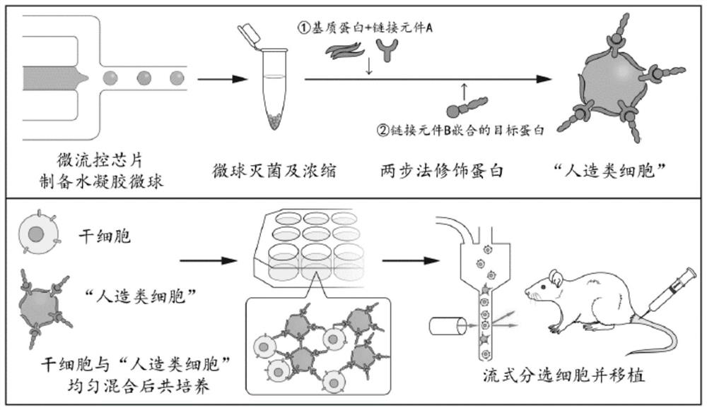
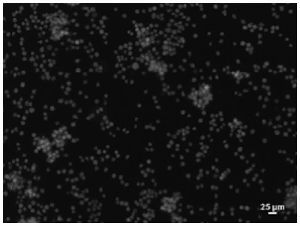
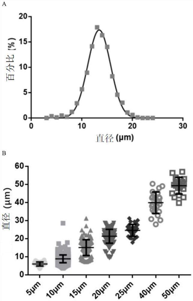
[0068] In this example, by simulating the shape and size of cells, hydrogel microspheres with a diameter of about 7-15 microns and a main material of polyethylene glycol-NAS were prepared for cell culture.

[0069] The preparation method of the hydrogel microspheres includes:

[0070] (1) Prepare a solution containing 10% polyethylene glycol diacrylate (PEGDA575), 0.1% N-acryloyloxysuccinimide (NAS) and 0.8% ammonium persulfate (APS) as a dispersed phase solution, 0.22um needle filter to remove impurities;

[0071] (2) The n-hexadecane solution containing 25% Span 80 (span80) was prepared as a continuous phase solution, and the 0.22um needle filter was filtered to remove impurities;

[0072] (3) Use two 10ml precision glass injectors to draw the same volume of continuous phase solution, and fix the two precision glass injectors into the Harvard precision dual-channel syringe pump slot; use one 1ml precision glass injector Draw an appropriate volume of disperse phase solution...

Embodiment 2

[0078] In this example, another hydrogel microsphere was prepared in a similar manner to Example 1. Compared with Example 1, the only difference is the use of 5% four-arm polyethylene glycol diacrylate (4-arm PEGAc), 0.5% dithiothreitol (DTT) and 0.1% N-acryloyloxysuccinate A solution of imide (NAS) was used as the dispersed phase solution, and a 25% span 80 in n-hexadecane solution was used as the continuous phase solution.

Embodiment 3

[0080] In this example, another hydrogel microsphere was prepared in a similar manner to Example 1. Compared with Example 1, the only difference is that an aqueous solution containing 3% gelatin (Gelatin) is used as the dispersed phase solution, and a n-hexadecane solution containing 25% span 80 (span80) is used as the continuous phase solution.


PUM

PropertyMeasurementUnit
diameteraaaaaaaaaa
diameteraaaaaaaaaa
elastic modulusaaaaaaaaaa

Abstract

The invention provides a protein modified microsphere and a method for culturing cells in vitro. The protein modified microspheres take hydrogel microspheres as a main body, and bioactive proteins interacting with cells, especially stem cells, are grafted on the surfaces of the hydrogel microspheres. The protein modified microspheres effectively simulate the interaction between supporting cells and stem cells in a natural microenvironment in a stem cell body, and stable and effective in-vitro expansion of the stem cells is realized.

Description

technical field [0001] The present disclosure relates to the field of biomedical engineering, in particular to a protein-modified microsphere and a method for culturing cells in vitro. Background technique [0002] In recent years, the cell therapy industry has developed rapidly, and the demand for in vitro culture of stem cells is soaring. [0003] In the related art, researchers introduced various cytokines such as stem cell factor, thrombopoietin, etc., and small molecules (such as UM171, SR1) into the in vitro culture of hematopoietic stem cells to promote the in vitro proliferation of hematopoietic stem cells. However, the maintenance or enhancement of reconstituted hematopoietic function after transplantation of hematopoietic stem cells in vitro remains unsatisfactory. Directly co-culturing the supporting cells in the natural microenvironment of stem cells with hematopoietic stem cells can improve the hematopoietic reconstruction effect of hematopoietic stem cells cul...

Claims


Application Information

Patent Timeline
Patent Type & Authority Applications(China)
IPC IPC(8): B01J13/00C12N5/0789
CPCB01J13/0065C12N5/0647C12N2533/52C12N2533/54C12N2533/56
Inventor 杜亚楠李文静敖艳肖梁海威李宁
Owner TSINGHUA UNIV
Learn More