
Cellular pathology image quality evaluation method

A technology for image quality evaluation and cytopathology, applied in image analysis, image data processing, neural learning methods, etc. The effect of accuracy

Pending Publication Date: 2022-07-29
HARBIN UNIV OF SCI & TECH
View PDF0 Cites 1 Cited by
  • Summary
  • Abstract
  • Description
  • Claims
  • Application Information

AI Technical Summary

Problems solved by technology

[0004] The purpose of the present invention is to solve the problem of the lack of a unified and standardized evaluation method for the quality of cytopathological images, and propose a method for evaluating the quality of cytopathological images

Method used


Image

  • Cellular pathology image quality evaluation method
  • Cellular pathology image quality evaluation method
  • Cellular pathology image quality evaluation method

Examples


Embodiment Construction

[0084] In order to make the purposes, technical solutions and advantages of the embodiments of the present invention clearer, the technical solutions in the embodiments of the present invention will be clearly and completely described below with reference to the accompanying drawings in the embodiments of the present invention. Obviously, the described embodiments These are some embodiments of the present invention, but not all embodiments. Based on the embodiments of the present invention, all other embodiments obtained by those of ordinary skill in the art without creative efforts shall fall within the protection scope of the present invention.

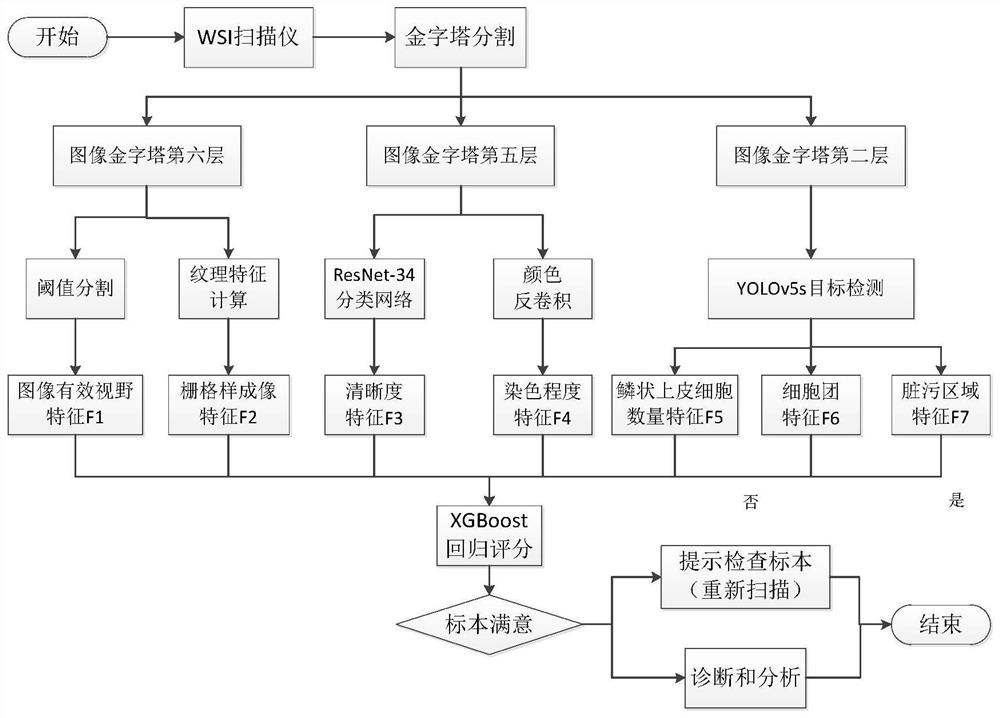
[0085] like figure 1 The implementation flow chart of the method in this paper is shown, including the steps:

[0086] 1. The scanner scans the sample to obtain a panoramic slice image. Then use the method of pyramid segmentation to obtain images of different layers of the pyramid;

[0087] 2. Using the image of the sixth layer o...


PUM


Abstract

The invention discloses a cell pathology image quality evaluation method, and relates to the problem of lack of a cell pathology image quality evaluation method in an automatic pathology diagnosis technology. The pathological section digitalization accumulates a large amount of data, which is an important premise of automatic pathological diagnosis. However, unqualified cell pathological images generated by scanning severely affect the efficiency of automatic pathological diagnosis, and even cause misdiagnosis and missed diagnosis. Many existing image quality evaluation methods do not consider evaluation standards of cell pathology doctors on sample images and are not suitable for the field of automatic pathological diagnosis. In order to solve the problem, the invention provides a cell pathology image quality evaluation method based on the Chinese expert consensus of digital pathology image acquisition and image quality control of cervical liquid-based cytology and the TBS (Theoresda system) diagnostic standard. Experiments show that the method can effectively evaluate the quality of the cell pathological image and improve the efficiency of automatic pathological diagnosis. The method is applied to quality evaluation of cell pathology images.

Description

technical field [0001] The present invention designs a cytopathological image quality evaluation method Background technique [0002] In recent years, the high incidence of cervical cancer has become a social problem that threatens women's lives. Among women worldwide, approximately 527,600 new cervical cancers are diagnosed each year and nearly 265,000 die from the disease, 90% of whom are located in underdeveloped areas. About 75,000 new cases of cervical cancer are discovered in China every year, accounting for 1 / 7 of the total number of new cases of cervical cancer in the world, and 35,000 people die from the disease. The current effective screening method for cervical cancer is the pathological examination of cervical exfoliated cell smear. With the advancement and development of science and technology, the traditional way of relying on pathologists to diagnose under a microscope faces many problems, such as time-consuming and labor-intensive, and it is greatly influe...

Claims


Application Information

Patent Timeline
IPC IPC(8): G06T7/00G06T7/10G06T7/62G06K9/62G06N3/04G06N3/08G06V10/82G06V10/762G06V10/764
CPCG06T7/0012G06T7/10G06T7/62G06N3/08G06T2207/30168G06T2207/20081G06T2207/20084G06T2207/30024G06T2207/30204G06T2207/30242G06N3/045G06F18/23G06F18/241
Inventor 何勇军刘皓晨王泽鑫赵晶谢怡宁
Owner HARBIN UNIV OF SCI & TECH
Learn More