


Quick Research
Generate reliable direction feasibility study reports for your R&D in just a few steps.

Technical Q&A
Discover and master advanced knowledge NOW. Basics, ideas, possibilities, all at once.

Find Solutions
As an expert in R&D theories, this can generate solutions to your technical problems instantly.

Evaluate Feasibility
Analyze your overall solution with one click, know your potential R&D risks in advance.

Monitor Landscape
Get weekly tech updates, stay abreast of the latest tech innovations and key insights.
Monitoring device for cell microdamage induction and bright field monitoring method
A monitoring device and cell technology are applied in the field of medical detection to achieve the effects of a reliable cell micro-damage induction method, simple equipment and simple structure
- Summary
- Abstract
- Description
- Claims
- Application Information
AI Technical Summary
Problems solved by technology
Method used
Image

Examples
Embodiment 1
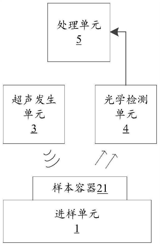
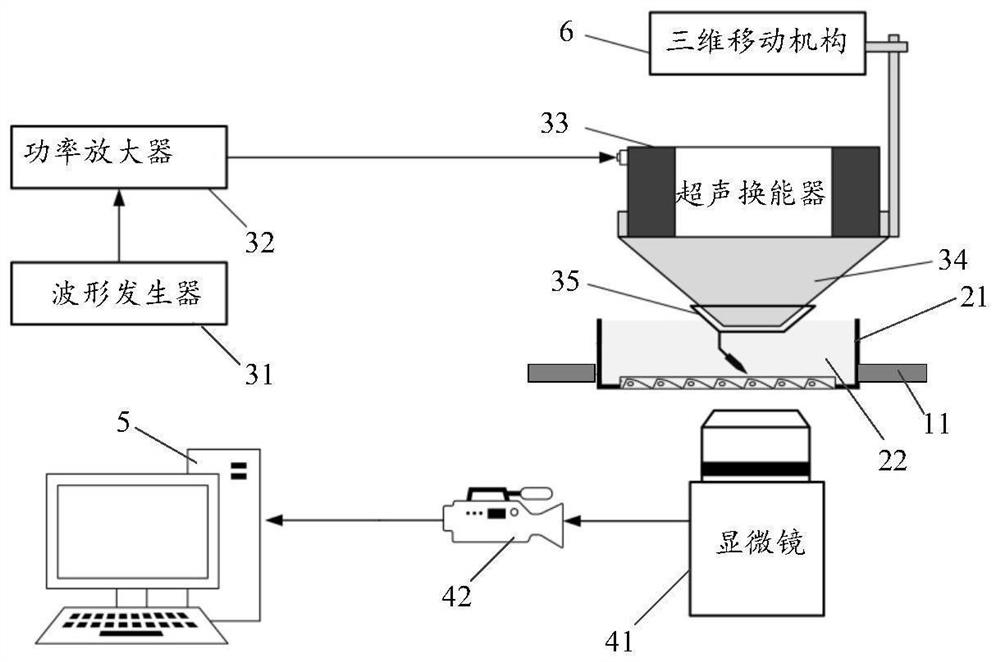
[0039] Please refer to figure 1 and figure 2 , this embodiment discloses a monitoring device for cell micro-damage induction, which mainly includes a sampling unit 1, an ultrasonic generating unit 3, an optical detection unit 4 and a processing unit 5, which will be described separately below.
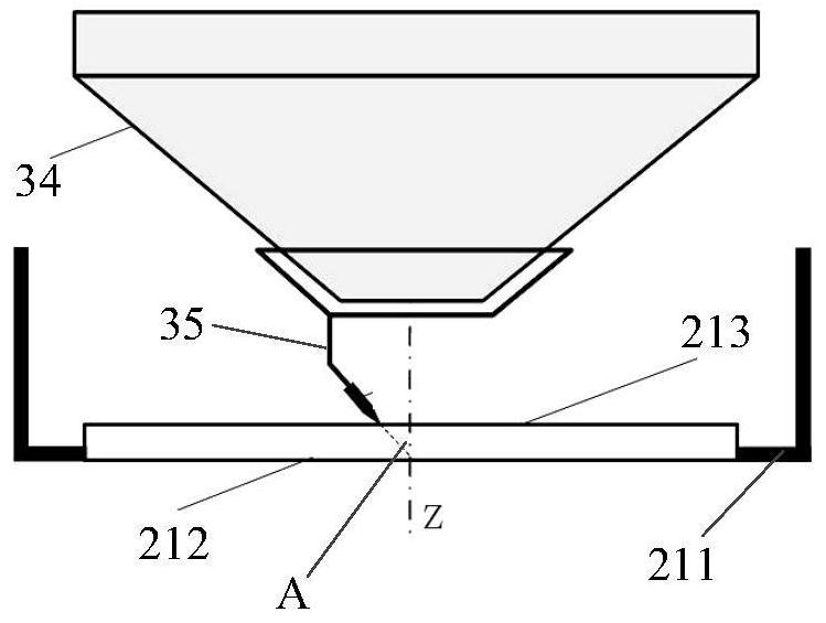
[0040] The sampling unit 1 has a detection table 11 for placing the sample container 21, and the detection table 11 may have a groove or a bracket adapted to the sample container 21, so that the sample container 21 can be placed stably. The sample container 21 can be a sample cup or a petri dish, which is used to accommodate the sample 22 to be tested formed by mixing the cell suspension solution and the cell microdamage solution. The sample 22 to be tested includes a plurality of cells and several microbubbles attached to each cell . Among them, the cell suspension is a solution of a certain concentration of living cells, which contains many living cells; the cell microinjury solut...
Embodiment 2
[0084] On the basis of the monitoring device disclosed in Example 1, this example discloses a bright field monitoring method for cell damage, the bright field monitoring method is mainly in figure 1 and figure 2 The application is carried out on the processing unit 5 in the
[0085] In this example, please refer to Figure 8 , the bright field monitoring method for cell micro-damage includes steps 110-160, which will be described respectively below.
[0086] Step 110, acquiring a bright-field image of micro-damaged cells in the sample to be tested.
[0087] For the monitoring device in Embodiment 1, see figure 1 and figure 2 , through the camera 42 in the optical detection unit 4, some images after the cells in the sample 22 to be tested are slightly damaged are captured, and a bright field image is formed and stored in the processing unit 5. Then, the processing unit 5 can obtain by reading One or more brightfield images. It should be noted that the bright field image...
Embodiment 3
[0105] On the basis of the bright field monitoring method for cell micro-damage disclosed in the second embodiment, a monitoring device is disclosed in this embodiment, and the monitoring device 7 includes a memory 71 and a processor 72 .
[0106] In this embodiment, the memory 71 and the processor 72 are the main components of the monitoring device 7. Of course, the monitoring device 7 may also include some detection components and execution components connected to the processor 72. For details, refer to the first embodiment above, here No more details.
[0107] Wherein, the memory 71 can be used as a computer-readable storage medium, and is used for storing a program here, and the program can be a program code corresponding to the bright field monitoring method in the second embodiment.
[0108] Wherein, the processor 72 is connected with the memory 71, and is used to execute the program stored in the memory 71 to realize the bright field monitoring method disclosed in the s...
PUM
| Property | Measurement | Unit |
|---|---|---|
| Diameter | aaaaa | aaaaa |
| Outer diameter | aaaaa | aaaaa |
| Inner diameter | aaaaa | aaaaa |
Abstract
Description
Claims
Application Information
 - R&D Engineer
- R&D Manager
- IP Professional
- Industry Leading Data Capabilities
- Powerful AI technology
- Patent DNA Extraction
Browse by: Latest US Patents, China's latest patents, Technical Efficacy Thesaurus, Application Domain, Technology Topic, Popular Technical Reports.
© 2024 PatSnap. All rights reserved.Legal|Privacy policy|Modern Slavery Act Transparency Statement|Sitemap|About US| Contact US: help@patsnap.com