
Image segmentation method and storage medium

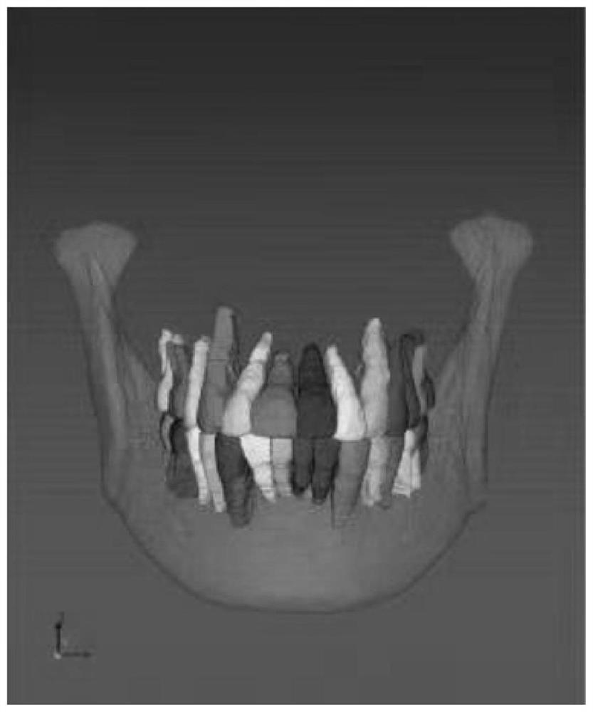
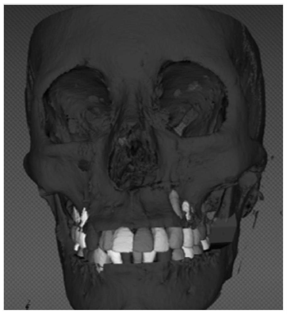
An image segmentation and image technology, applied in the field of medical image processing, can solve the problems of low efficiency and low accuracy of manual segmentation of tooth graphics, and achieve the effect of improving the efficiency and accuracy of diagnosis and treatment, reducing consumption time, and shortening segmentation time.

Inactive Publication Date: 2022-03-11
SHANGHAI XUHUI DISTRICT DENTAL CENT +1
View PDF0 Cites 1 Cited by
  • Summary
  • Abstract
  • Description
  • Claims
  • Application Information

AI Technical Summary

Problems solved by technology

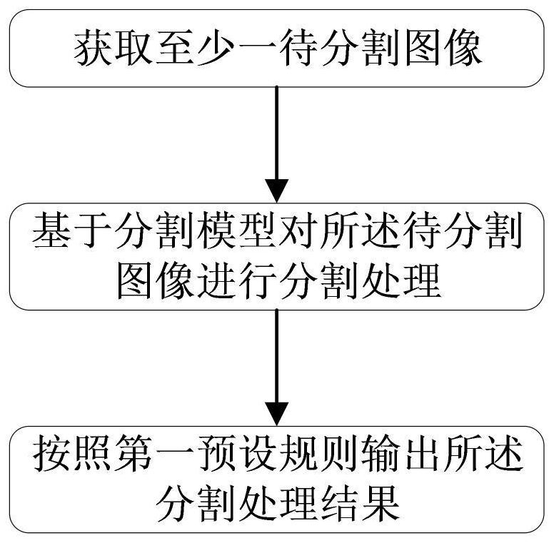
[0006] In order to solve the technical problems of low efficiency and low accuracy in manual segmentation of tooth graphics in the prior art, the present application aims to provide an image segmentation method and storage medium, which can improve segmentation accuracy while improving segmentation efficiency

Method used


Image

  • Image segmentation method and storage medium
  • Image segmentation method and storage medium
  • Image segmentation method and storage medium

Examples


Embodiment Construction

[0049] Reference will now be made in detail to the exemplary embodiments, examples of which are illustrated in the accompanying drawings. When the following description refers to the accompanying drawings, the same numerals in different drawings refer to the same or similar elements unless otherwise indicated. The implementations described in the following exemplary embodiments do not represent all implementations consistent with this application. Rather, they are merely examples of apparatuses and methods consistent with aspects of the present application as recited in the appended claims.

[0050] It should be noted that, in this document, the term "comprising", "comprising" or any other variation thereof is intended to cover a non-exclusive inclusion such that a process, method, article or apparatus comprising a set of elements includes not only those elements, It also includes other elements not expressly listed, or elements inherent in the process, method, article, or de...


PUM


Abstract

The invention discloses an image segmentation method. The method comprises the following steps: acquiring at least one to-be-segmented image; performing segmentation processing on the to-be-segmented image based on a segmentation model; and outputting the segmentation processing result according to a first preset rule. Through the image segmentation method provided by the invention, the segmentation model can be continuously trained according to the segmentation case and data, so that the accuracy of the segmentation model is higher and higher, the time consumed by a doctor in segmenting the tooth image can be effectively reduced through the high-accuracy segmentation model, the segmentation time of the tooth image is shortened, and the segmentation efficiency of the tooth image is improved. And the diagnosis and treatment efficiency and accuracy are improved.

Description

technical field [0001] The present application relates to the technical field of medical image processing, in particular to an image segmentation method and a storage medium. Background technique [0002] At present, the specific method of using CT for 3D reconstruction is as follows: import the included CBCT in the form of dicom data into mimics software (Materialize Co, Leuven, Belgium), and manually separate bone tissue and teeth. During the separation process, the dentist manually marks the jawbone and the target tooth with different colors, and manually sets different transparency to show each structure and its relationship with other surrounding structures. [0003] Due to the similar density of bones and teeth in the above operations, hundreds of CT levels need to be distinguished by the physician himself based on experience. If the image quality is not good, there are artifacts, etc., further adjustments are required. This process is complicated, and experienced de...

Claims


Application Information

Patent Timeline
Patent Type & Authority Applications(China)
IPC IPC(8): G06T7/00G06T7/10
CPCG06T7/0012G06T7/10G06T2207/10081G06T2207/20081G06T2207/20084G06T2207/30036
Inventor 钱文昊胡易瑞
Owner SHANGHAI XUHUI DISTRICT DENTAL CENT
Learn More