Medical image three-dimensional reconstruction method based on voxel growth

A medical image and three-dimensional reconstruction technology, applied in the field of medical images, can solve the problems of unfavorable organ structures and overall display of lesions in tomographic images, and achieve the effects of accelerating traversal speed, reducing calculations, and avoiding ambiguity problems

Pending Publication Date: 2022-01-04
HARBIN UNIV OF SCI & TECH
View PDF0 Cites 0 Cited by
  • Summary
  • Abstract
  • Description
  • Claims
  • Application Information

AI Technical Summary

Problems solved by technology

However, tomographic images are not conducive to the overall display of organ structures and lesions. Continuous observation of multiple frames of images, integration of human brain thinking or the use of image post-processing and reconstruction techniques, can form a complete concept

Method used


Image

  • Medical image three-dimensional reconstruction method based on voxel growth
  • Medical image three-dimensional reconstruction method based on voxel growth
  • Medical image three-dimensional reconstruction method based on voxel growth

Examples


Embodiment Construction

[0021] In order to make the features of the present invention clearer, the present invention will be further described below in conjunction with the accompanying drawings and examples.

[0022] The present invention is a three-dimensional reconstruction method of medical images based on voxel growth. Taking the patient's coccyx as an example, the image after preprocessing and segmentation is used as the read-in data for three-dimensional reconstruction, and the method of the present invention is used to reconstruct and extract equivalent values. noodle.

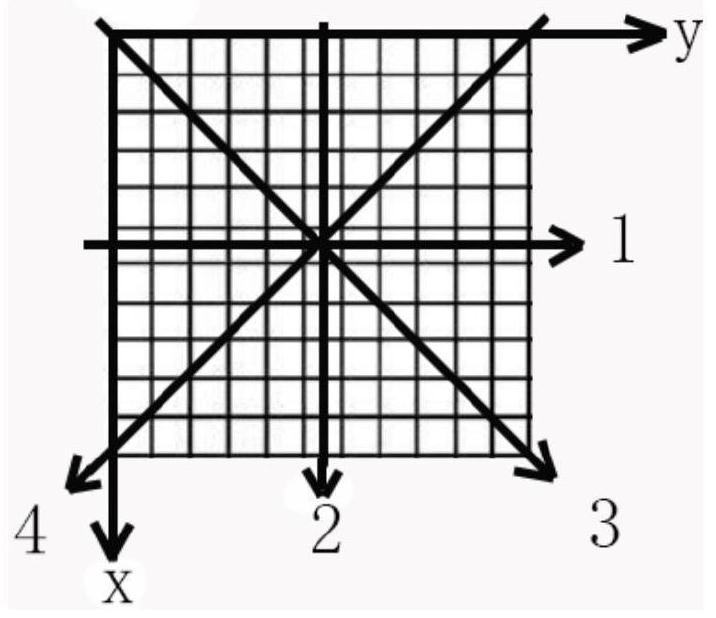
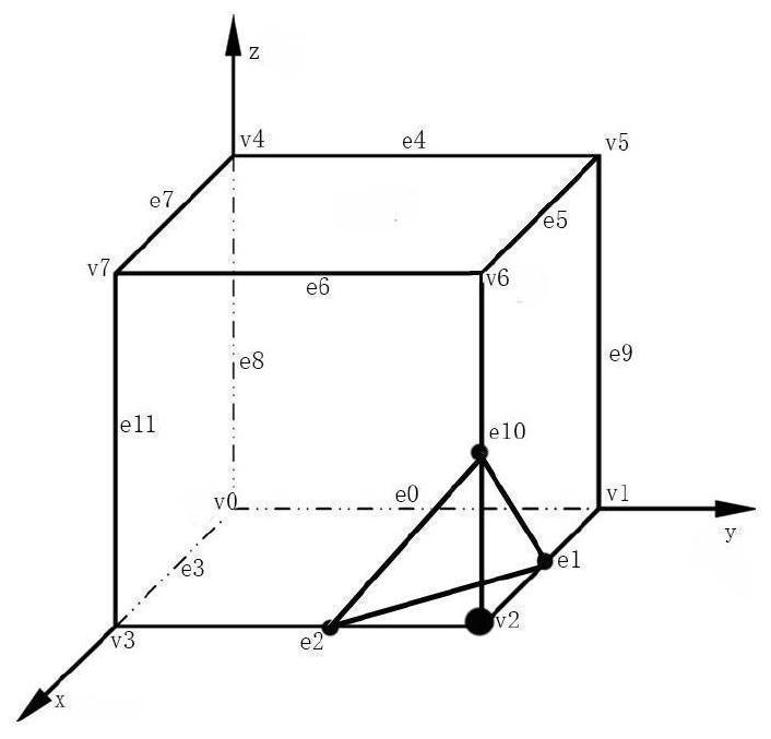
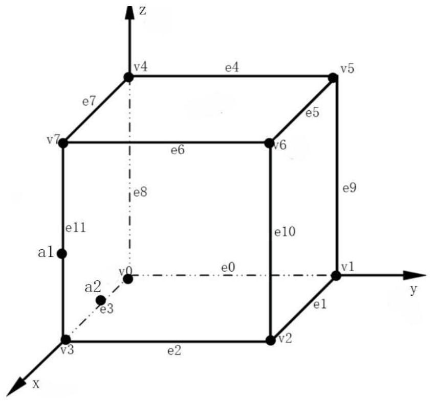
[0023] Firstly, create the extended configuration triangular index table 2 and the growth index table 3 for the extended configuration. And the face index values ​​of the six growth directions and the interpolation point index tables corresponding to the six directions established according to the constructed cube information, the order of front, back, left, right, up, down, respectively is: Index_f=v 2 v 3 v 7 v 6 , Inde...


PUM


Abstract

The invention belongs to the technical field of medical images, and provides a medical image three-dimensional reconstruction contour surface extraction method. According to the method, two index tables of an original MC algorithm need to be improved firstly, and then a new growth index table, surface index values in six growth directions and corresponding six surface equivalent point index tables are created. Firstly, a middle layer of cube is traversed in a shape like a Chinese character'mi 'to find seed voxels, and then the seed voxels are used for growth reconstruction. In the increasing process, index values in six directions and an equivalent point index table are used for ensuring the construction of adjacent cube information, that is, equivalent point information solved on a common surface and adjacent voxel information are put into an increasing queue together, so that the same equivalent point information does not need to be repeatedly calculated when adjacent voxels are reconstructed, wherein a problem of ambiguity is effectively avoided by adopting an expansion lookup table method. Then, a growth lookup table is constructed to determine the growth direction, so that traversal of cubes without contour surfaces is avoided, and the reconstruction time is shortened. The steps are repeated, and the contour surface reconstruction is ended when the queue is empty.

Description

technical field [0001] The invention belongs to the field of medical images, in particular to a method for extracting isosurface grids of three-dimensional reconstruction of medical images. Background technique [0002] With the development of medical imaging technology, medical imaging has become an important means for doctors to judge diseases. They can clearly display the tissue structure of various organs and significantly improve the detection rate of lesions. However, tomographic images are not conducive to the overall display of organ structures and lesions. It is necessary to continuously observe multiple frames of images, integrate human brain thinking or use image post-processing and reconstruction technology to form a complete concept. Using medical two-dimensional tomographic images to reconstruct and display three-dimensional images of human tissues, it greatly improves the doctor's diagnosis rate of diseases. At present, the most mature medical image grid gene...

Claims


Application Information

Patent Timeline
Patent Type & Authority Applications(China)
IPC IPC(8): G06T17/00
CPCG06T17/00G06T2210/41
Inventor 王沫楠王鑫夏领兵
Owner HARBIN UNIV OF SCI & TECH
Why Eureka
Try Eureka