
Ultrasonic detection device for ophthalmology department

An ultrasonic detection and ophthalmology technology, applied in eye examination, ultrasonic/sonic/infrasonic diagnosis, structure of ultrasonic/sonic/infrasonic diagnostic equipment, etc., can solve problems such as burying diseases and restricting the final effect

Pending Publication Date: 2021-12-21
上海百杨视光技术有限公司
View PDF0 Cites 0 Cited by
  • Summary
  • Abstract
  • Description
  • Claims
  • Application Information

AI Technical Summary

Problems solved by technology

Errors often restrict the final effect, and there will be a phenomenon of burying the disease

Method used


Image

  • Ultrasonic detection device for ophthalmology department
  • Ultrasonic detection device for ophthalmology department
  • Ultrasonic detection device for ophthalmology department

Examples


Embodiment Construction

[0026] Embodiments of the present invention are described in detail below, examples of which are shown in the drawings, wherein the same or similar reference numerals designate the same or similar elements or elements having the same or similar functions throughout. The embodiments described below by referring to the figures are exemplary and are intended to explain the present invention and should not be construed as limiting the present invention.

[0027] In describing the present invention, it should be understood that the terms "center", "longitudinal", "transverse", "length", "width", "thickness", "upper", "lower", "front", " The orientations or positional relationships indicated by "rear", "left", "right", "vertical", "horizontal", "top", "bottom", "inner", "clockwise", "counterclockwise" are based on the attached The orientation or positional relationship shown in the figure is only for the convenience of describing the present invention and simplifying the description...


PUM


Abstract

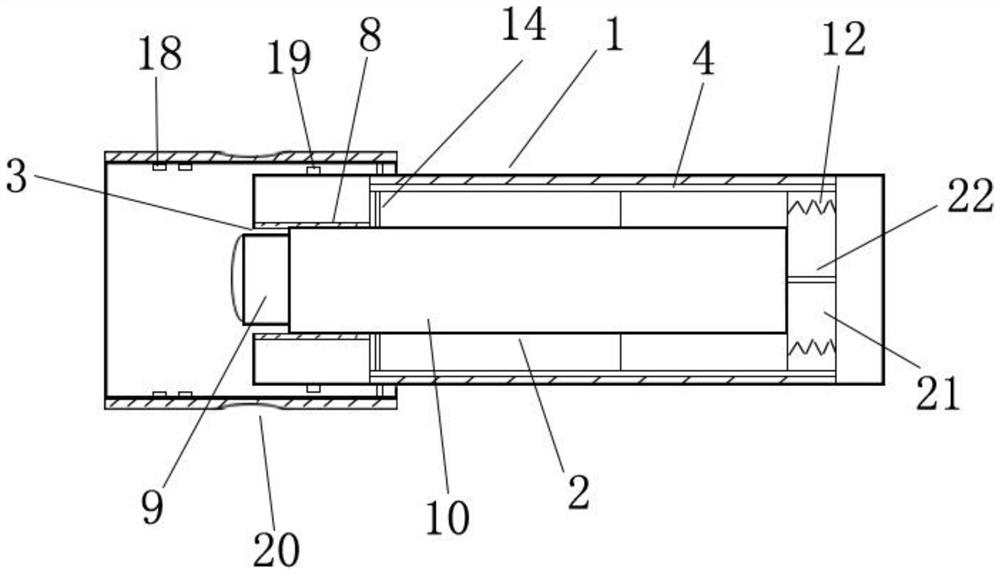
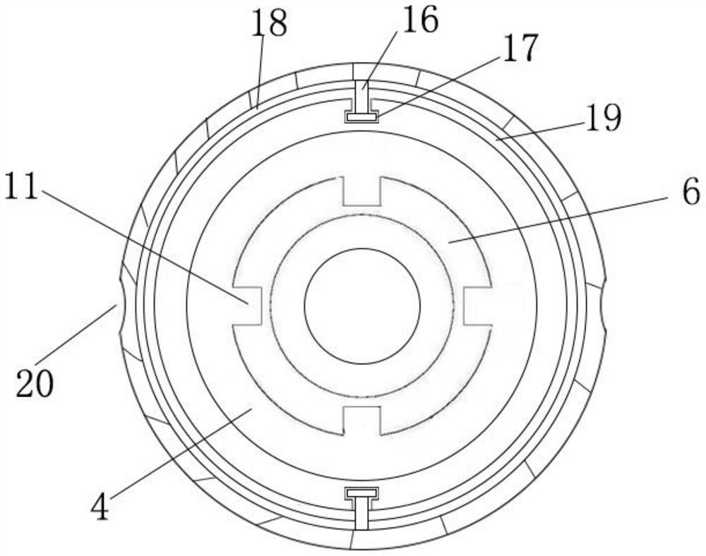
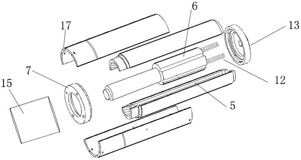
The invention relates to the field of ultrasonic detection equipment, in particular to an ultrasonic detection device for the ophthalmology department. The ultrasonic detection device comprises a shell, and the shell comprises an outer shell body and a probe part; a cavity is formed in the outer shell body, and the probe part can be axially and movably arranged in the cavity; a through hole allowing the probe part to pass through is formed in the front end of the outer shell body, and when the front end of the probe part is flush with the front end of the outer shell body, a gap exists between the rear end of the probe part and the rear wall of the cavity; the front end of the probe part can extend out of the through hole under the action of self gravity; and after extending out of the through hole and making contact with the cornea of the eyeball, the probe part can retract into the outer shell body by means of the elastic force of the cornea of the eyeball. According to the technical scheme, the probe part can naturally fall and extend out of the end part of the outer shell body under the gravity of the probe part, can automatically retract when being subjected to a certain external force, and can automatically retract when being subjected to corneal elasticity when being in contact with eyes during ophthalmic examination; and the probe part is hardly subjected to pressure during use, so that the detection error is reduced.

Description

technical field [0001] The invention relates to the field of ultrasonic detection equipment, in particular to an ophthalmic ultrasonic detection device. Background technique [0002] A-ultrasound is the abbreviation of A-type ultrasound. It is based on the relationship between the time and amplitude of the sound wave to detect the echo of the sound wave, and its positioning accuracy is high. For ophthalmic A-ultrasound, the probe is placed in front of the eyes, and the sound beam propagates forward. Every time a reflection occurs at an interface, the echoes are arranged on the baseline in the form of peaks according to the return time, and the echo intensity is represented by the height of the peak. The higher the value, the formation of a one-dimensional linear image, the higher the tissue discrimination. It can display the anterior chamber depth, crystal thickness, vitreous cavity length and axial length with an accuracy of 0.01mm, which is used for eye structure measurem...

Claims


Application Information

Patent Timeline
Patent Type & Authority Applications(China)
IPC IPC(8): A61B8/10
CPCA61B8/10A61B8/44A61B8/4444
Inventor 不公告发明人
Owner 上海百杨视光技术有限公司
Learn More