
Multi-information fused pathological image classification and recognition method and system

A pathological image, classification and recognition technology, applied in the field of medical imaging diagnosis, can solve the problems of classification method lag, incomplete utilization, and insufficient accuracy of histopathological image classification, and achieve the effect of improving diagnostic effect and efficiency

Pending Publication Date: 2021-12-14
上海派影医疗科技有限公司
View PDF2 Cites 3 Cited by
  • Summary
  • Abstract
  • Description
  • Claims
  • Application Information

AI Technical Summary

Problems solved by technology

The above methods all use deep learning automatic recognition as the technical basis, but there are still deficiencies in the accuracy of histopathological image classification, and there is a certain lag in the classification method based on full-section histopathological images
In addition, in the process of image diagnosis, the advantages of computer aids are not fully utilized, and there is a lack of auxiliary functions with users in business logic, and there is still room for research and development in the provision of diagnostic information.

Method used


Image

  • Multi-information fused pathological image classification and recognition method and system
  • Multi-information fused pathological image classification and recognition method and system
  • Multi-information fused pathological image classification and recognition method and system

Examples


Embodiment Construction

[0067] The application will be further described in detail below in conjunction with the accompanying drawings and embodiments. It should be understood that the specific embodiments described here are only used to explain related inventions, rather than to limit the invention. It should also be noted that, for the convenience of description, only the parts related to the related invention are shown in the drawings.

[0068] It should be noted that, in the case of no conflict, the embodiments in the present application and the features in the embodiments can be combined with each other. The present application will be described in detail below with reference to the accompanying drawings and embodiments.

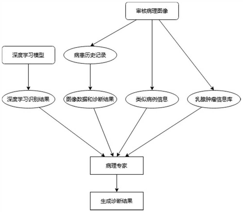
[0069] figure 1 An exemplary system architecture 100 of a pathological image classification and recognition method fused with multiple information according to the embodiment of the present application is shown.

[0070] Such as figure 1 As shown, the system architecture 1...


PUM


Abstract

The invention provides a multi-information fused pathological image classification and recognition method and system. The method comprises the steps of pathological data management, pathological image preprocessing, pathological image recognition, cancerization area prediction and diagnosis report generation. The system is divided into a background management server and a pathology expert terminal. The management background server is used for expert personal information storage management, patient data storage and pathology image data management. The pathology expert terminal is responsible for hospital experts to upload pathology images and patient information, and classification and recognition of the pathology images are realized through a pre-trained deep neural network model. A pathological expert can audit a deep learning recognition result through the expert terminal and generate a diagnosis report. According to the invention, pathological conditions of the pathological images are judged and a tumor area is predicted through a deep learning method, a benign tumor result and a malignant tumor result are automatically predicted, the area where the tumor is located is visually displayed on the pathological images, a doctor is assisted in diagnosing the pathological images, and the diagnosis effect and efficiency are improved.

Description

technical field [0001] The invention relates to the technical field of medical image diagnosis, in particular to a pathological image classification and recognition method and system that integrates multiple information. Background technique [0002] Breast cancer is the most common form of cancer in women. In 2017 alone, studies show that approximately 252,000 new invasive breast cancers and 63,000 in situ breast cancers are expected to be diagnosed, and 2,000 breast cancer-related deaths are expected to occur. Therefore, it is necessary to carry out early diagnosis and treatment to reduce the incidence rate and improve the quality of life of patients. Histopathology remains the key to the diagnostic process and is the gold standard for distinguishing between benign and malignant tissues, and between patients with carcinoma in situ and invasive carcinoma. Diagnosis and differentiation of breast cancer subtypes usually involves the collection of tissue biopsies from masses...

Claims


Application Information

Patent Timeline
IPC IPC(8): G06T7/00G06K9/32G06K9/62G06N3/04G06N3/08G16H50/20
CPCG06T7/0012G06N3/08G16H50/20G06T2207/20104G06T2207/20081G06T2207/30068G06T2207/30096G06N3/045G06F18/241
Inventor 郑魁丁维龙朱筱婕赵樱莉李涛余鋆
Owner 上海派影医疗科技有限公司
Learn More