
Biological analysis method for detecting concentration of human VEGF-A (vascular endothelial growth factor-A) by using chemical luminescence method

A VEGF-A technology using chemistry, applied in the field of biological analysis and detection, can solve the problems of high instrument prices, high kit costs, and high sensitivity requirements for detection methods, and achieve low reagent costs, low cost, and high sensitivity.

Pending Publication Date: 2021-10-22
宁波熙宁检测技术有限公司 +1
View PDF7 Cites 1 Cited by
  • Summary
  • Abstract
  • Description
  • Claims
  • Application Information

AI Technical Summary

Problems solved by technology

Due to the low concentration of VEGF-A in the plasma sample anticoagulated with sodium citrate, the sensitivity of the detection method is high, and the detection limit of conventional ELISA kits such as R&Dsystems Cat.DVE00 in serum plasma is 31.3pg / mL, unable to meet the needs of detection
The detection limit of the electrochemiluminescence detection method MSD Cat.K151UVK in serum and plasma is 2pg / mL, which can meet the demand, but the cost of the whole kit is very high, and the price of the supporting equipment is high. Compared with ELISA kits, there are many choices. The MSD electrochemiluminescence detection method has a monopoly position, and there are no alternative products to choose from. Once the procurement cycle is too long or out of stock, it is not conducive to the continuation of the clinical detection business of VEGF-A concentration determination.

Method used


Image

  • Biological analysis method for detecting concentration of human VEGF-A (vascular endothelial growth factor-A) by using chemical luminescence method
  • Biological analysis method for detecting concentration of human VEGF-A (vascular endothelial growth factor-A) by using chemical luminescence method
  • Biological analysis method for detecting concentration of human VEGF-A (vascular endothelial growth factor-A) by using chemical luminescence method

Examples


Embodiment 1

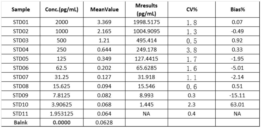
[0024] The establishment of embodiment 1 ELISA method VEGFA standard curve

[0025] Anti-VEGF fusion protein (Baiying Biotechnology, Cat. No. B576801) was diluted to 2 μg / mL with 1XPBS, 100 μL / well was added to 96 microplate plate (Shenzhen Jincanhua Industrial, 106-004), and incubated overnight for 4 hours. The next day, discard the microplate solution, wash three times with washing solution (washing solution component 1×PBS+0.1% Tween 20), pat dry, add 300 μL / well blocking solution (blocking solution / method analysis solution component 1 ×PBS+1%BSA+0.1%Tween 20), incubate at room temperature 200rpm for 1 hour; VEGFA standard (the standard product comes from the kit R&D product number DY293B-05) uses the sample analysis solution (the composition of the sample diluent is the method analysis solution, add 10 % rabbit serum) for gradient dilution, the dilution initial concentration is 500pg / mL, then two-fold gradient dilution 10 points, the standard concentration range of the sta...

Embodiment 2

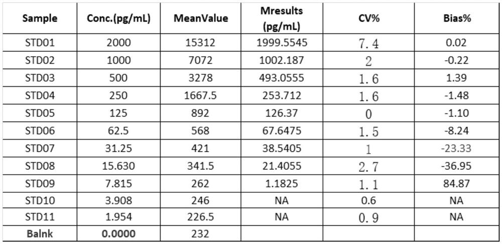
[0028] The establishment of embodiment 2 MSD method VEGFA standard curve

[0029] Anti-VEGF fusion protein (Baiying Biotechnology, Cat. No. B576801) was diluted to 2 μg / mL with 1XPBS, 100 μL / well was added to the MSD plate (MSD, Cat. No. L15XA-3), and incubated overnight at 4. The next day, discard the microplate solution, wash three times with washing solution (washing solution component 1×PBS+0.1% Tween 20), pat dry, add 300 μL / well blocking solution (blocking solution / method analysis solution component 1 ×PBS+1%BSA+0.1%Tween 20), incubate at room temperature 200rpm for 1 hour; VEGFA standard ((standard product comes from the kit R&D product number DY293B-05) using the sample analysis solution (the composition of the sample diluent is the method analysis solution added 10% rabbit serum) for serial dilution, the dilution initial concentration is 500pg / mL, then two-fold serial dilution 10 points, the standard concentration range of the standard curve is 2000pg / mL, 1000pg / mL, 5...

Embodiment 3

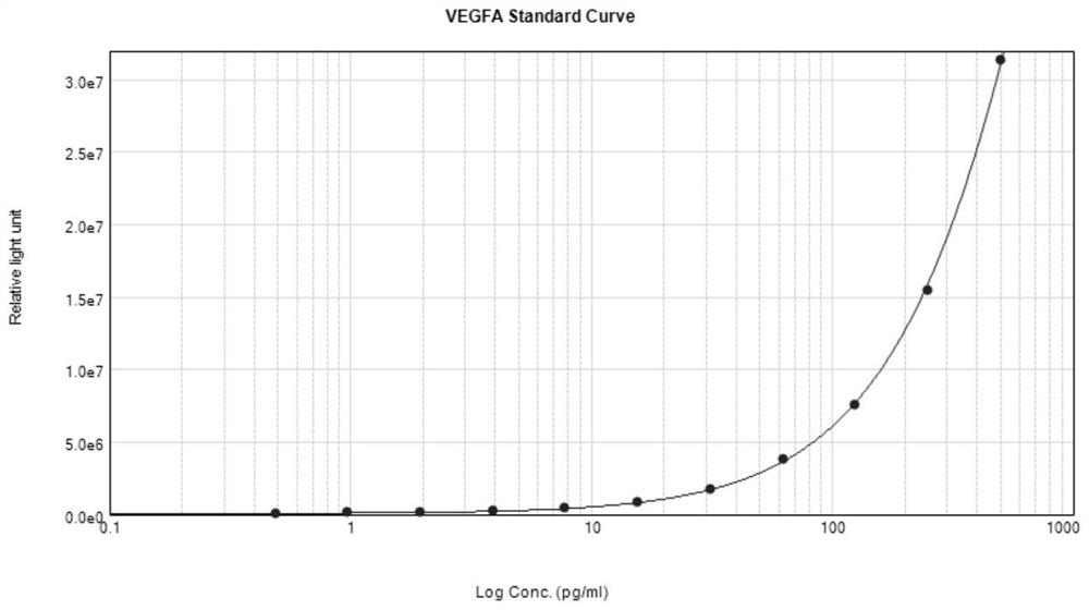
[0032] The establishment of embodiment 3 chemiluminescence method VEGFA standard curve

[0033] The anti-VEGF fusion protein (Baiying Biotechnology, Cat. No. B576801) was diluted to 2 μg / mL with 1X PBS, 100 μL / well was added to a 96 microplate white plate (thermo 436110), and incubated overnight for 4 hours. The next day, discard the microplate solution, wash three times with washing solution (washing solution component 1×PBS+0.1% Tween 20), pat dry, add 300 μL / well blocking solution (blocking solution / method analysis solution component 1 ×PBS+1%BSA+0.1%Tween 20), incubate at room temperature 200rpm for 1 hour; VEGFA standard ((standard product comes from the kit R&D product number DY293B-05) using the sample analysis solution (the composition of the sample analysis solution is added to the method analysis solution) 10% rabbit serum) for gradient dilution, the dilution initial concentration is 500pg / mL, then two-fold gradient dilution 10 points, the standard concentration rang...


PUM


Abstract

The invention discloses a biological analysis method for detecting the concentration of human VEGF-A. On the basis of a ligand-receptor binding principle, a coating protein, a detection substance VEGFA and a detection antibody are bound on a solid medium in sequence, and then a chromogenic substrate is added for detection, wherein the method comprises the following steps: a specific protein of VEGF-A is fixed by using a solid medium, namely coating protein, a detection sample is added, the VEGF-A in the sample is specifically fixed on the solid medium, and a second antibody, namely a detection antibody, which can be combined with the VEGF-A is fixed on the fixing medium, The non-specifically adsorbed impurities are washed away along with the washing process; the second antibody needs to be directly or indirectly specially marked, and finally, a substrate corresponding to the marker is used for color development detection. The detection sensitivity is high, the cost is relatively low, the source is controllable, and clinical sample analysis can be continuously carried out.

Description

technical field [0001] The invention relates to the field of biological analysis and detection, in particular to a method for detecting the concentration of VEGF-A in a human matrix by means of a double-antibody sandwich method and a chemiluminescence detection method. Background technique [0002] Vascular endothelial growth factor (VEGF) is a cytokine that plays an important role in the process of angiogenesis. The VEGF family includes VEGF-A, VEGF-B, VEGF-C, VEGF-D and VEGF-E, as well as placental growth factor (PIGF). VEGF-A has diverse physiological activities, including promoting proliferation and migration of endothelial cells, promoting proteolysis, and promoting microvascular leakage, all of which promote angiogenesis. VEGF-A exerts physiological effects by binding VEGFR-1 and VEGFR-2. Hypoxia induces VEGF-A expression under inflammatory conditions and glucose deprivation. VEGF-A expression is upregulated in inflamed and vascularized corneas. VEGF-A also plays a...

Claims


Application Information

Patent Timeline
Patent Type & Authority Applications(China)
IPC IPC(8): G01N33/68G01N33/58G01N33/574G01N33/543G01N21/76
CPCG01N33/6872G01N33/6893G01N33/74G01N33/581G01N33/57488G01N33/54393G01N21/76G01N2333/475G01N2800/16G01N2800/52
Inventor 黄启宽朱国振余跃云胡国清刘野杨雨生
Owner 宁波熙宁检测技术有限公司
Learn More