Cell nucleus segmentation and counting method and system for immunohistochemical cell images
A technology of immunohistochemistry and counting methods, which is applied in image analysis, image data processing, image enhancement and other directions, and can solve the problems of large amount of calculation, insufficient accuracy, and irregular nuclei of stained cells in deep learning algorithms.
- Summary
- Abstract
- Description
- Claims
- Application Information
AI Technical Summary
Problems solved by technology
Method used
Image
Examples
Embodiment 1
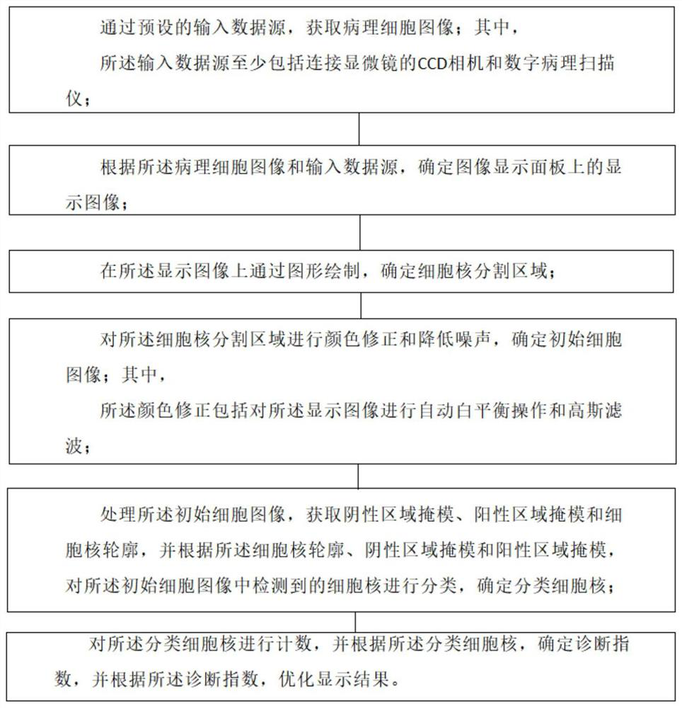
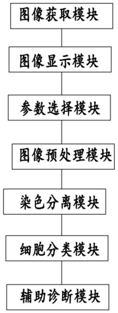
[0104] according to Figure 1-9 As shown, the present invention provides a method for segmenting and counting cell nuclei in immunohistochemical cell images, which is characterized in that it includes:
[0105] Obtain pathological cell images through preset input data sources; where,
[0106] The input data source includes at least a CCD camera connected to a microscope and a digital pathology scanner;
[0107] Determine the display image on the image display panel according to the pathological cell image and the input data source;
[0108] Determining the segmented area of the cell nucleus by graphically drawing on the displayed image;
[0109] Carrying out color correction and noise reduction on the cell nucleus segmentation area to determine the initial cell image; wherein,
[0110] The color correction includes performing automatic white balance operation and Gaussian filtering on the displayed image;
[0111] Processing the initial cell image to obtain a negative ar...
Embodiment 2
[0116] according to Figure 1-9 As shown, the technical solution provides an embodiment, the determination of the display image on the image display panel according to the pathological cell image and the input data source further includes:
[0117] When the input data source is a CCD camera connected to a microscope, the pathological cell image is updated in real time based on the preset CCD camera sampling, and the display image is determined;
[0118] When the input data source is a digital pathology scanner, based on the user's needs, perform area selection on the cell image to determine the display image; wherein,
[0119] The region selection includes at least zooming and panning.
[0120] The working principle and beneficial effects of the above-mentioned technical scheme are:
[0121] The input data source of this technical solution can be a CCD camera connected to a microscope, or a digital pathological image obtained by a digital pathology scanner. In the CCD camera...
Embodiment 3
[0123] according to Figure 1-9 As shown, the technical solution provides an embodiment,
[0124] The step of determining the cell nucleus segmented area by graphically drawing on the displayed image includes:
[0125] Determining the area of the cell nucleus based on the image display panel;
[0126] According to the area of the nucleus, a graphic frame is drawn for the nucleus to determine the division area of the nucleus; wherein,
[0127] The graphic frame includes a rectangle, a circle or an arbitrary curve.
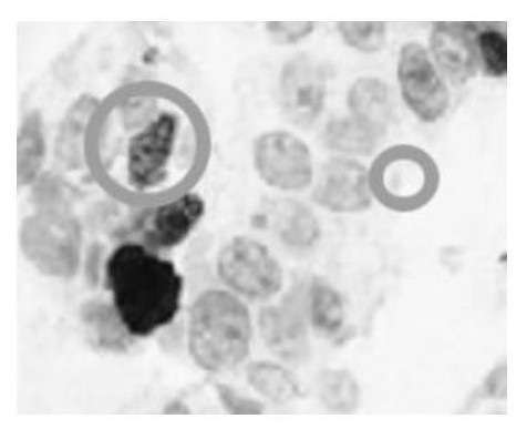
[0128] In actual implementation, the present invention also adopts the display area segmented according to the graphic frame and the cell to determine the segmentation parameters; wherein,
[0129] The segmentation parameters include circular parameters and rectangular parameters; wherein,
[0130] The circle parameter is used to set the minimum area of the nucleus and the color threshold of the positive level;
[0131] The rectangle parameter is used t...
PUM
 Abstract
Description
Claims
Application Information
 - Generate Ideas
- Intellectual Property
- Life Sciences
- Materials
- Tech Scout
- Unparalleled Data Quality
- Higher Quality Content
- 60% Fewer Hallucinations
Browse by: Latest US Patents, China's latest patents, Technical Efficacy Thesaurus, Application Domain, Technology Topic, Popular Technical Reports.
© 2025 PatSnap. All rights reserved.Legal|Privacy policy|Modern Slavery Act Transparency Statement|Sitemap|About US| Contact US: help@patsnap.com


