Jugular vein ball socket and sinus sigmoideus groove positioning method and intelligent temporal bone image processing system

A jugular bulb and sigmoid sulcus technology, applied in the field of medical image processing, can solve the problems of complex adjacent structures, high error rate and high time complexity, and achieve the effect of easy implementation and promotion

Active Publication Date: 2021-08-06
BEIJING FRIENDSHIP HOSPITAL CAPITAL MEDICAL UNIV +1
View PDF3 Cites 0 Cited by
  • Summary
  • Abstract
  • Description
  • Claims
  • Application Information

AI Technical Summary

Problems solved by technology

When the perivascular bone wall associated with pulsatile tinnitus is missing, the lesion site is changeable, the imaging manifestations are different, and the surrounding structures are adjacent to each other, which becomes a difficult point in imaging diagnosis, which brings challenges to its detection and localization
[0005]The conventional target detection method uses a sliding window to detect in an entire image during the positioning process, and the time complexity is high; the anatomical structure changes with the continuous layer The sequence will change in shape, and it is difficult for conventional object detection methods to classify the changing objects into the same category, so the error rate is usually high when detecting continuously changing anatomical structures; conventional object detection methods usually require a large amount of labeled data for Model training, which consumes a lot of manpower and time

Method used


Image

  • Jugular vein ball socket and sinus sigmoideus groove positioning method and intelligent temporal bone image processing system
  • Jugular vein ball socket and sinus sigmoideus groove positioning method and intelligent temporal bone image processing system
  • Jugular vein ball socket and sinus sigmoideus groove positioning method and intelligent temporal bone image processing system

Examples


Embodiment Construction

[0042] Exemplary embodiments of the present invention will be described in more detail below with reference to the accompanying drawings. Although exemplary embodiments of the present invention are shown in the drawings, it should be understood that the invention may be embodied in various forms and should not be limited to the embodiments set forth herein. Rather, these embodiments are provided for more thorough understanding of the present invention and to fully convey the scope of the present invention to those skilled in the art.

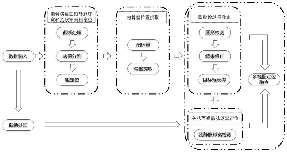
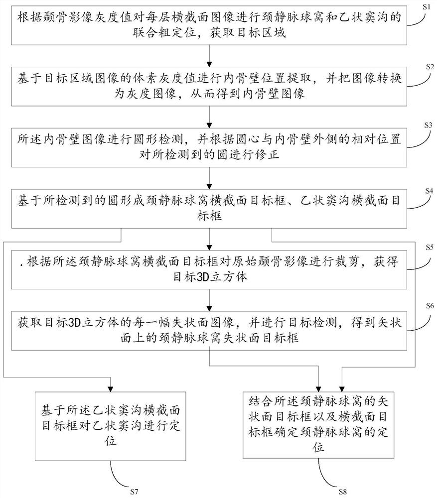
[0043] The present invention fully considers the positional relationship between different layers of anatomical structures in medical images and the shape characteristics of each anatomical structure itself, and considers the characteristics of 3D data, and fully utilizes multi-view information to improve the accuracy of detection and positioning. The present invention proposes a method for automatically positioning the jugular socket and the si...


PUM


Abstract

The invention provides an automatic positioning method for a jugular vein ball socket and a sinus sigmoideus groove in a temporal bone image and an intelligent temporal bone image processing system, which are used for computer-assisted pulsatile tinnitus diagnosis. According to the method, a thick-to-thin frame is adopted to carry out step-by-step accurate positioning, and the shape information and relative position information of the jugular vein ball socket and the sinus sigmoideus groove are fully utilized, so that the detection and positioning algorithm is more efficient and lighter. By combining and fusing multi-view information, the positioning effect is optimized, and the detection accuracy of a multi-layer continuous change target is improved. Compared with a deep learning method, the automatic positioning method provided by the invention does not depend on a large amount of labeled data any more, and is easier to implement and popularize.

Description

technical field [0001] The invention relates to the field of medical image processing, in particular to a jugular ball socket and sigmoid groove positioning method and an intelligent temporal bone image processing system. Background technique [0002] Tinnitus is manifested as the abnormal perception of sound by the human body without external stimuli. It is a common and frequently-occurring disease in otology. Among them, pulsatile tinnitus (Pulsatile Tinnitus, PT) accounts for about 4%-10% of tinnitus patients. In the past ten years, with economic development and people's emphasis on health, the demand for diagnosis and treatment of patients has surged. With the popularization of temporal bone imaging technology, the imaging method for pulsatile tinnitus has become an important technical means. [0003] Temporal bone CT is the main method for diagnosing osseous lesions in pulsatile tinnitus. Temporal bone CT can display multiple fine structures such as the middle ear and...

Claims


Application Information

Patent Timeline
Patent Type & Authority Applications(China)
IPC IPC(8): G06T7/00G06T7/13G06T7/136G06T7/181G06N3/04G06N3/08
CPCG06T7/0012G06T7/136G06T7/13G06T7/181G06N3/08G06T2207/10081G06T2207/20081G06T2207/30008G06N3/045
Inventor 王振常周祎超李晓光尹红霞卓力赵鹏飞吕晗
Owner BEIJING FRIENDSHIP HOSPITAL CAPITAL MEDICAL UNIV
Why Eureka
Try Eureka