
Method and system for precise registration and coincidence of preoperative CT or nuclear magnetic image and corresponding focus in operation, storage medium, and equipment

A technology for accurate registration of nuclear magnetic images, applied in image analysis, image enhancement, image data processing, etc. Narrow and small problems, to achieve the effect of improving deformability, solving deformation, and solving interference factors

Active Publication Date: 2021-06-04
沈阳蓝软智能医疗科技有限公司 +1
View PDF5 Cites 3 Cited by
  • Summary
  • Abstract
  • Description
  • Claims
  • Application Information

AI Technical Summary

Problems solved by technology

[0003] However, at present, the clinical application of 3D medical imaging is only suitable for preoperative surgical planning, and has not given full play to its important role in intraoperative navigation, especially in clinical operations related to non-rigid solid organs and tissues such as liver and kidney. Guidance, because most of these organs involve minimally invasive surgery, in the real surgical environment, the endoscope used in minimally invasive surgery has narrow field of view, large noise and soft tissue deformation during operation during the process of collecting endoscopic images. These factors seriously affect the registration accuracy of the patient's 3D model and the lesion area, so that the registration accuracy cannot meet the requirements of intraoperative navigation

Method used


Image

  • Method and system for precise registration and coincidence of preoperative CT or nuclear magnetic image and corresponding focus in operation, storage medium, and equipment
  • Method and system for precise registration and coincidence of preoperative CT or nuclear magnetic image and corresponding focus in operation, storage medium, and equipment
  • Method and system for precise registration and coincidence of preoperative CT or nuclear magnetic image and corresponding focus in operation, storage medium, and equipment

Examples


Embodiment Construction

[0109] The present invention will be described in detail below with reference to the accompanying drawings and examples.

[0110] In order to make the purpose, technical solutions and advantages of the embodiments of the present invention clearer, the technical solutions in the embodiments of the present invention will be clearly and completely described below in conjunction with the drawings in the embodiments of the present invention. Obviously, the described embodiments It is only some embodiments of the present invention, but not all embodiments. The following description of at least one exemplary embodiment is merely illustrative in nature and in no way taken as limiting the invention, its application or uses. Based on the embodiments of the present invention, all other embodiments obtained by persons of ordinary skill in the art without making creative efforts belong to the protection scope of the present invention.

[0111] The non-rigid solid organs and tissues involv...


PUM


Abstract

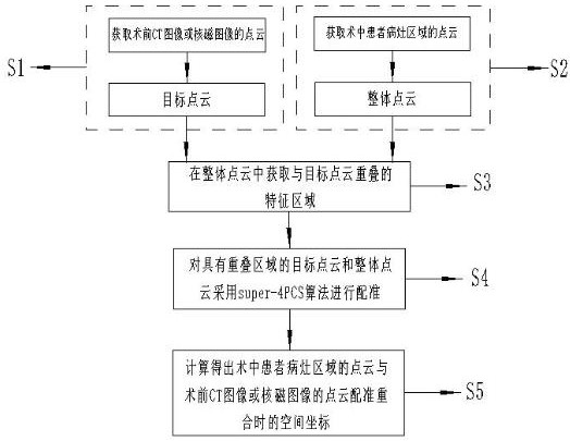
The invention relates to a method for precise registration and coincidence of a preoperative CT or nuclear magnetic image and a corresponding focus in an operation. The method is used for assisting planning and implementation of a clinical operation. The method comprises the following steps: acquiring a point cloud of a preoperative CT image or a nuclear magnetic image as a target point cloud; acquiring a point cloud of a focus area of a patient in an operation as an integral point cloud; acquiring a feature region overlapped with the target point cloud from the overall point cloud; adopting a super-4PCS algorithm to carry out registration on the target point cloud and the overall point cloud with the overlapped areas, and obtaining point cloud coordinates of the focus area of the patient in the operation, wherein the point cloud coordinates are space coordinates when the point cloud of the focus area of the patient in the operation and the point cloud of the preoperative CT image or the nuclear magnetic image are registered and coincide. According to the method, the registration precision problem of the three-dimensional model of the non-rigid organs and tissues of the patient and the focus area of the patient in the real operation environment is mainly solved, a doctor can quickly and accurately find the organ corresponding to the three-dimensional model of the patient during the operation, and the three-dimensional model is accurately matched with the organ.

Description

technical field [0001] The present invention relates to a method for precise registration and coincidence of preoperative CT or nuclear magnetic images and corresponding intraoperative lesions, which is used to assist the planning and implementation of clinical operations, especially the planning and operation related to organs and tissues of non-rigid entities. implement. Background technique [0002] Three-dimensional medical imaging is more and more commonly used in auxiliary clinical operations. It is a 3D model file generated after three-dimensional reconstruction based on the patient's CT or MRI sequence images. Reality technology, presented in the augmented reality wearable device, can be used as a "panoramic map" of the doctor's surgical object before or during the operation, allowing the doctor to see the situation of the patient's lesion area or the preoperative plan at a glance, thus replacing During the traditional operation, the doctor observes the CT or MRI pl...

Claims


Application Information

Patent Timeline
IPC IPC(8): G06T7/33G06T7/11G06T7/00G06N3/04G06N3/08
CPCG06T7/33G06T7/0012G06T7/11G06N3/08G06T2207/10028G06T2207/10081G06T2207/20081G06T2207/20084G06T2207/30084G06T2207/30096G06N3/045
Inventor 杨云鹏谢锦华孙野
Owner 沈阳蓝软智能医疗科技有限公司
Learn More