Dual-wavelength enhanced Raman endoscopic non-invasive pathological detection device and detection method

A Raman endoscopic and enhanced technology, applied in the fields of endoscopy, diagnostic recording/measurement, medical science, etc., can solve the problems of difficult to achieve pathological diagnosis, difficult to obtain Raman spectrum, increase the risk of use, etc. Accurate and reliable non-invasive histopathological diagnosis, enhanced Raman spectral signal intensity, stable autofocus and zoom measurement effects

Active Publication Date: 2021-06-01
TSINGHUA UNIV
View PDF5 Cites 5 Cited by
  • Summary
  • Abstract
  • Description
  • Claims
  • Application Information

AI Technical Summary

Problems solved by technology

[0004] In the existing research, the existing research on the combination of Raman spectroscopy and endoscopy is to achieve in-situ real-time Raman spectroscopy measurement through the combination of fiber optic Raman and endoscopy, without considering the impact of the internal environment on Raman spectroscopy. Due to the complex influence of spectral signals, the lack of reliable and high-precision Raman scattering signal measurement methods, and the lack of feasible pathological analysis methods based on Raman spectral signals, it is not yet possible to truly achieve non-invasive pathological detection
Some studies have further improved the Raman endoscope, but in the actual measurement of biological tissues, there are very obvious fluorescent signals, which seriously affect the measurement signal-to-noise ratio, peak position, and peak intensity fitting accuracy of Raman spectroscopy.
At the same time, the Raman signal intensity of the sample itself is weak, and increasing the integration time can increase the intensity of the measured signal to a certain extent, but if the Raman integration time is blindly increased, the detection time will become longer, which will undoubtedly increase the pain of the patient
In the field of Raman measurement, the surface-enhanced Raman method and the tip-enhanced Raman method can effectively increase the Raman signal intensity of the sample to be measured, but this technology has not been applied to the field of Raman endoscopy
[0005] On the other hand, the existing research does not consider the focusing problem in living tissue, and the focusing effect will directly affect the measurement accuracy of Raman
Existing endoscopes often use an auxiliary focus ring to help focus, but on the one hand, applying the focus ring will increase the diameter of the endoscope lens and increase the patient’s discomfort during use; on the other hand, the focus ring may fall during operation, increasing the In order to avoid the risk of use, it is urgent to design an integrated self-focusing device
At the same time, the endoscope structure designed by some methods cannot be coupled with the existing clinical endoscope, and may even increase the size of the endoscope, which may increase the suffering of patients in practical applications, and it is difficult to obtain Raman spectra that can be used effectively , difficult to achieve pathological diagnosis
In addition, the Raman method for three-dimensional scanning pathological analysis has not been proposed, and it is difficult to determine the scope and depth of the lesion
[0006] In summary, the existing Raman endoscopic measurement methods are still unable to measure Raman spectra that eliminate the influence of fluorescence and have better signal strength
At the same time, the existing Raman endoscope device is still unable to realize the three-dimensional scanning Raman measurement of the integrated self-focusing and zooming of the lens.
In addition, after obtaining high-quality Raman spectra, there is still a lack of reliable pathological analysis criteria extraction methods and spectral information comparison methods, so as to truly realize the pathological detection of tissues.

Method used


Image

  • Dual-wavelength enhanced Raman endoscopic non-invasive pathological detection device and detection method
  • Dual-wavelength enhanced Raman endoscopic non-invasive pathological detection device and detection method
  • Dual-wavelength enhanced Raman endoscopic non-invasive pathological detection device and detection method

Examples


Embodiment 1

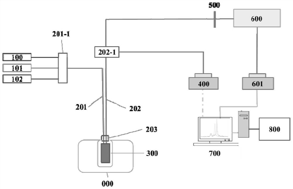
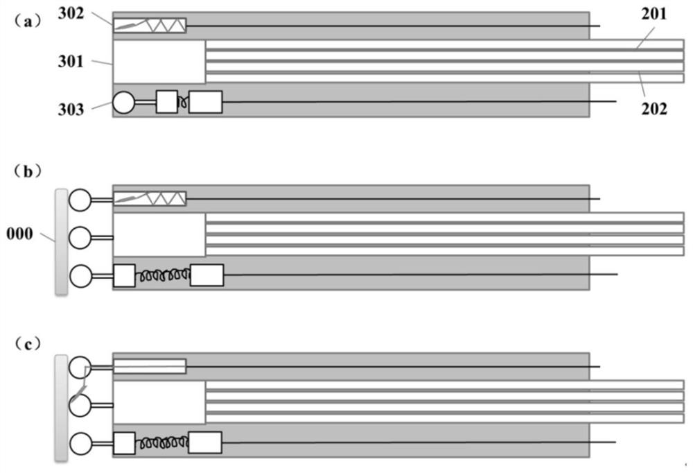
[0082] In this embodiment, the structure of the endoscopic lens 300 is as Figure 5 As shown in (a), it includes an objective lens 301 , a signal enhancement device 302 , a retractable support foot 303 and a channel exit 304 . In the existing commercial endoscope structure, the objective lens 301 and the channel exit 304 are inherent structures, and the telescopic support feet 303 can be embedded into the endoscope lens through simple modification. substitute. It is only necessary to lead the designed signal enhancement device 302 to the endoscope lens through the channel outlet 304, and the dual-wavelength enhanced self-focusing Raman endoscope measurement can be realized based on the method proposed by the present invention.

[0083] At this point, the measurement process is:

[0084] 1) Insert the endoscope lens 300 connected with the optical fiber bundle to the position to be measured 000, the three supporting feet 303 (or auxiliary focus ring) on ​​the endoscope lens ar...

Embodiment 2

[0095] In this embodiment, the structure of the endoscopic lens 300 is as Figure 5 As shown in (b), it includes an objective lens 301 , a signal enhancement device 302 , and a telescopic support foot 303 . Since there is no need to judge the pathological information by sampling biopsy, for the first-time patients, it is not necessary to use the endoscopic lens with the clamp channel, and Figure 5 (a) By comparison, it is easy to find that when other components have the same size, the diameter of the endoscope designed in the second embodiment can be greatly reduced compared with the diameter of the existing commercial endoscope, which significantly reduces the pain of the patient being examined.

[0096] Then the measurement process at this time is compared with Embodiment 1, only the measurement process 3-1) needs to be replaced by:

[0097] 3-1) Turn on the signal enhancement device 302 to realize enhanced Raman spectrum measurement. The specific opening methods include,...


PUM


Abstract

The invention discloses a dual-wavelength enhanced Raman endoscopic non-invasive pathological detection device and a detection method, which are combined with endoscope detection, Raman detection and pathological analysis to directly detect a Raman spectrum of tissues in a living body and perform non-invasive pathological analysis based on an artificial intelligence method through spectral information to replace traditional invasive pathological examination. By constructing a signal enhancement device, the measured Raman spectrum signal is enhanced, and the nondestructive pathological analysis of the in-vivo tissue is effectively realized. Two kinds of laser with different wavelengths are used for detecting tissue signals of the same part, the difference value is calculated through the two sets of signals, the fluorescence influence is eliminated, and the measurement signal-to-noise ratio of the Raman endoscope is increased. The endoscope lens of a self-focusing structure is designed, self-focusing and zooming of the endoscope are achieved on the basis that the diameter of the endoscope lens is not increased through built-in supporting legs, and three-dimensional scanning pathological detection of a sample is achieved on the basis that a more stable and clearer spectrum result is obtained. Through adoption of an artificial intelligence pathology analysis method, non-invasive pathology detection is effectively realized.

Description

technical field [0001] The invention relates to the technical field of Raman measurement, in particular to a dual-wavelength enhanced Raman endoscopic non-invasive pathological detection device and detection method. Background technique [0002] Cancer has become one of the major public health problems that seriously threaten the life and health of the Chinese population. In recent years, the incidence and mortality of cancer in my country have continued to rise, while the cancer survival rate has a large gap compared with developed countries. One of the main reasons for the above situation is the low diagnosis rate of early stage cancer. At present, for gastric cancer, esophageal cancer, nasopharyngeal cancer and other cancers, the most important diagnostic methods are endoscopic diagnosis and pathological biopsy of lesion tissue taken out of the body, but it has many limitations, especially in early cancer diagnosis. obvious. The endoscopic detection system is mainly a t...

Claims


Application Information

Patent Timeline
Patent Type & Authority Applications(China)
IPC IPC(8): A61B1/06A61B1/07A61B1/04A61B1/005A61B5/00
CPCA61B1/0661A61B1/07A61B1/04A61B1/005A61B1/00188A61B1/00186A61B5/0075
Inventor 张兴樊傲然张晓宇马维刚王海东
Owner TSINGHUA UNIV
Why Eureka
Try Eureka