
Fundus retinal blood vessel image processing system and method

A blood vessel image and processing system technology, applied in the field of fundus retina blood vessel image processing system, can solve the problems of low accuracy and complicated operation, and achieve the effect of qualitative analysis and quantitative analysis

Active Publication Date: 2021-05-28
THE FIRST AFFILIATED HOSPITAL OF HENAN UNIV OF SCI & TECH
View PDF6 Cites 0 Cited by
  • Summary
  • Abstract
  • Description
  • Claims
  • Application Information

AI Technical Summary

Problems solved by technology

[0003] However, the methods for processing fundus images in the prior art are more complex and less accurate. Therefore, there is an urgent need for a technical solution with simple operation and high accuracy in this field.

Method used


Image

  • Fundus retinal blood vessel image processing system and method
  • Fundus retinal blood vessel image processing system and method
  • Fundus retinal blood vessel image processing system and method

Examples


Embodiment 1

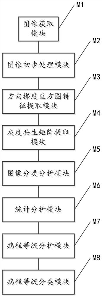
[0085] see figure 1 , a fundus blood vessel image processing system, specifically comprising:

[0086] The image acquisition module M1 is used to acquire fundus blood vessel images;

[0087] The image preliminary processing module M2 is used to perform preliminary processing on the fundus blood vessel image to obtain the preliminary processed fundus blood vessel image. This process specifically includes:

[0088] Extract the green image of the fundus blood vessel image; since the original image is an RGB image, and the image contrast of the green channel is the highest, the green image of the RGB image is extracted.

[0089] Performing histogram equalization processing on the green image to obtain the preliminarily processed fundus blood vessel image with blood vessel enhancement; performing histogram equalization processing on the image of the green channel to enhance the blood vessels in the image.

[0090] The histogram of oriented gradient feature extraction module M3 is...

Embodiment 2

[0178] see Figure 7 , an embodiment of the present invention provides a fundus blood vessel image processing method, including:

[0179] S1. Obtain fundus blood vessel images;

[0180] S2. Preliminary processing is performed on the fundus blood vessel image to obtain a preliminary processed fundus blood vessel image;

[0181] S3. Extracting the directional gradient histogram feature of the preliminary processed fundus blood vessel image;

[0182] S4. Extracting the gray level co-occurrence matrix of the preliminary processed fundus vascular image;

[0183] S5. Using the directional gradient histogram feature and the gray level co-occurrence matrix as an input of the SVM, perform image classification analysis, and obtain a classification analysis result.

[0184] As an optional implementation manner, in the embodiment of the present invention, preliminary processing is performed on the fundus blood vessel image, and after the preliminary processing of the fundus blood vesse...


PUM


Abstract

The invention relates to a fundus blood vessel image processing system and method, and the system comprises an image obtaining module, an image preliminary processing module, a histogram of oriented gradient feature extraction module, a gray level co-occurrence matrix extraction module, and an image classification analysis module. Through segmentation, feature extraction and classification of fundus images in the database, qualitative analysis and quantitative analysis of the image are realized, and the diameter, the blood vessel included angle, the fractal dimension and the tortuosity of the image after primary processing are calculated so that the image is predicted and classified more effectively.

Description

technical field [0001] The invention relates to the field of image processing, in particular to a system and method for image processing of fundus retinal blood vessels. Background technique [0002] Automatically extracting vascular target parameters from the background of fundus images is a basic task, but it is difficult to automatically extract vascular target parameters from fundus images with low resolution or uneven brightness changes. At present, many methods for processing fundus images have been proposed, such as methods based on threshold, based on mathematical morphology, based on feature extraction and based on machine learning. [0003] However, the methods for processing fundus images in the prior art are relatively complicated to operate and have low accuracy. Therefore, a technical solution with simple operation and high accuracy is urgently needed in this field. Contents of the invention [0004] The object of the present invention is to provide a system...

Claims


Application Information

Patent Timeline
Patent Type & Authority Applications(China)
IPC IPC(8): G06T7/00G06T7/11G06T7/13G06T7/136G06K9/38G06K9/46G06K9/62
CPCG06T7/0012G06T7/11G06T7/136G06T7/13G06T2207/10024G06T2207/20081G06T2207/20101G06T2207/20156G06T2207/30041G06T2207/30101G06V10/28G06V10/56G06V10/50G06F18/2411
Inventor 白永杰许梦莹白娇红李振伟杨晓利
Owner THE FIRST AFFILIATED HOSPITAL OF HENAN UNIV OF SCI & TECH
Learn More