A kind of polypeptide imaging probe and its preparation method and application
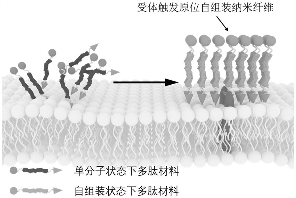
A technology for imaging probes and peptides, which is applied in the fields of polypeptide imaging probes and their preparation, in situ self-assembly polypeptide imaging probes for identifying cell receptors and their preparation, and can solve the problem of large molecular weight probes and limited imaging probes. The scope of application, the inability to enter efficiently, etc., to achieve the effect of enhancing the imaging signal-to-noise ratio, good aggregation retention ability, and simple structure
- Summary
- Abstract
- Description
- Claims
- Application Information
AI Technical Summary
Problems solved by technology
Method used
Image
Examples
Embodiment 1
[0073] Example 1 Design, synthesis and functional identification of polypeptide imaging probe αvβ3-Cy
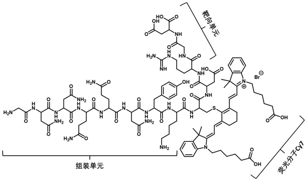
[0074] In this example, a polypeptide imaging probe αvβ3-Cy is designed with αVβ3 as the targeting receptor. The polypeptide imaging probe αvβ3-Cy is composed of a αVβ3 receptor recognition peptide, a soluble self-assembling peptide and a near-infrared fluorescent molecule Cy. The near-infrared fluorescent molecule Cy is connected to the side chain of the polypeptide through cysteine (C), and the molecular structure is as follows figure 2 As shown, the amino acid sequence is shown in SEQ ID NO: 14;
[0075] SEQ ID NO: 14: GNNQQNYKC(Cy7)DRGD.
[0076] The synthesis steps are as follows:
[0077] (1) Weigh the resin and put it into the peptide solid-phase synthesis tube, add an appropriate amount of DMF to swell for more than 4 hours; extract the DMF, use the Fmoc deprotection agent for Fmoc deprotection, and mix on a shaker for 15 minutes; extract the Fmoc deprotection a...
Embodiment 2
[0087] Example 2 Design, synthesis and functional identification of polypeptide imaging probe EpCAM-Cy
[0088] In this example, a polypeptide imaging probe EpCAM-Cy is designed with EpCAM as the targeting receptor. The polypeptide imaging probe EpCAM-Cy is composed of EpCAM receptor recognition peptide, soluble self-assembling peptide and near-infrared fluorescent molecule Cy. The near-infrared fluorescent molecule Cy is connected to the side chain of the polypeptide through cysteine (C), and the molecular structure is as follows Figure 5 As shown, the amino acid sequence is shown in SEQ ID NO: 15;
[0089] SEQ ID NO: 15: GNNQQNYKC(Cy7)DYEVHTYYLD.
[0090] The synthetic method is shown in Example 1.
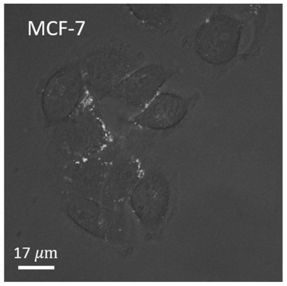
[0091] to about 10 5 Breast cancer cell lines MCF-7 and 10 with high expression of epithelial cell adhesion molecule EpCAM 5 1 mL of 20 μM polypeptide imaging probe EpCAM-Cy was added to HUVEC, a human umbilical vein endothelial cell line with low expression of the epithe...
Embodiment 3
[0096] Example 3 Design, synthesis and functional identification of polypeptide imaging probe αvβ3-NBD
[0097] Compared with near-infrared fluorescent molecules, the in vivo imaging effect of short-wavelength fluorescent molecules is slightly insufficient, but the imaging effect of cells is more stable and not easy to be quenched. In this example, NBD, a molecule with aggregation-induced luminescence effect, is used instead of the near-infrared fluorescent molecule Cy7 to perform fluorescence imaging at the cell level, which has the advantages of no washing, high signal-to-noise ratio, and can qualitatively characterize the expression of cell surface receptors.
[0098] The molecular structural formula of the polypeptide imaging probe αvβ3-NBD in this example is as follows: Figure 8 As shown, the amino acid sequence is as shown in SEQID NO:16, wherein X is Fmoc aminovaleric acid;
[0099] SEQ ID NO: 16: NBD-X-GNNQQNYRGD.
[0100] The synthesis method is shown in Example 1....
PUM
 Abstract
Description
Claims
Application Information
 - R&D
- Intellectual Property
- Life Sciences
- Materials
- Tech Scout
- Unparalleled Data Quality
- Higher Quality Content
- 60% Fewer Hallucinations
Browse by: Latest US Patents, China's latest patents, Technical Efficacy Thesaurus, Application Domain, Technology Topic, Popular Technical Reports.
© 2025 PatSnap. All rights reserved.Legal|Privacy policy|Modern Slavery Act Transparency Statement|Sitemap|About US| Contact US: help@patsnap.com


