
CT image artifact removing method

A CT image and image technology, applied in the field of medical imaging, can solve problems such as blurring and loss of image details, and achieve the effect of simple implementation method, low consumption, and preservation of image details

Active Publication Date: 2021-05-11
FMI MEDICAL SYST CO LTD
View PDF5 Cites 0 Cited by
  • Summary
  • Abstract
  • Description
  • Claims
  • Application Information

AI Technical Summary

Problems solved by technology

The disadvantage of this method is that it will cause the image details to be lost and become more blurred

Method used


Image

  • CT image artifact removing method
  • CT image artifact removing method
  • CT image artifact removing method

Examples


Embodiment Construction

[0035] In order to make the technical means of the present invention and the technical effects that can be achieved more clearly and more perfectly disclosed, an embodiment is provided hereby, and the following detailed description is given in conjunction with the accompanying drawings:

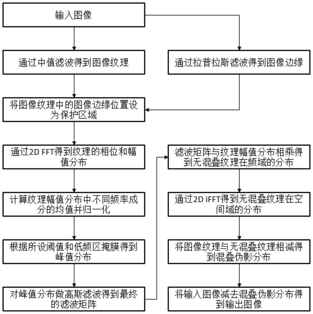
[0036] Such as figure 1 As shown, a method for removing artifacts from a CT image in this embodiment is implemented through the following steps:

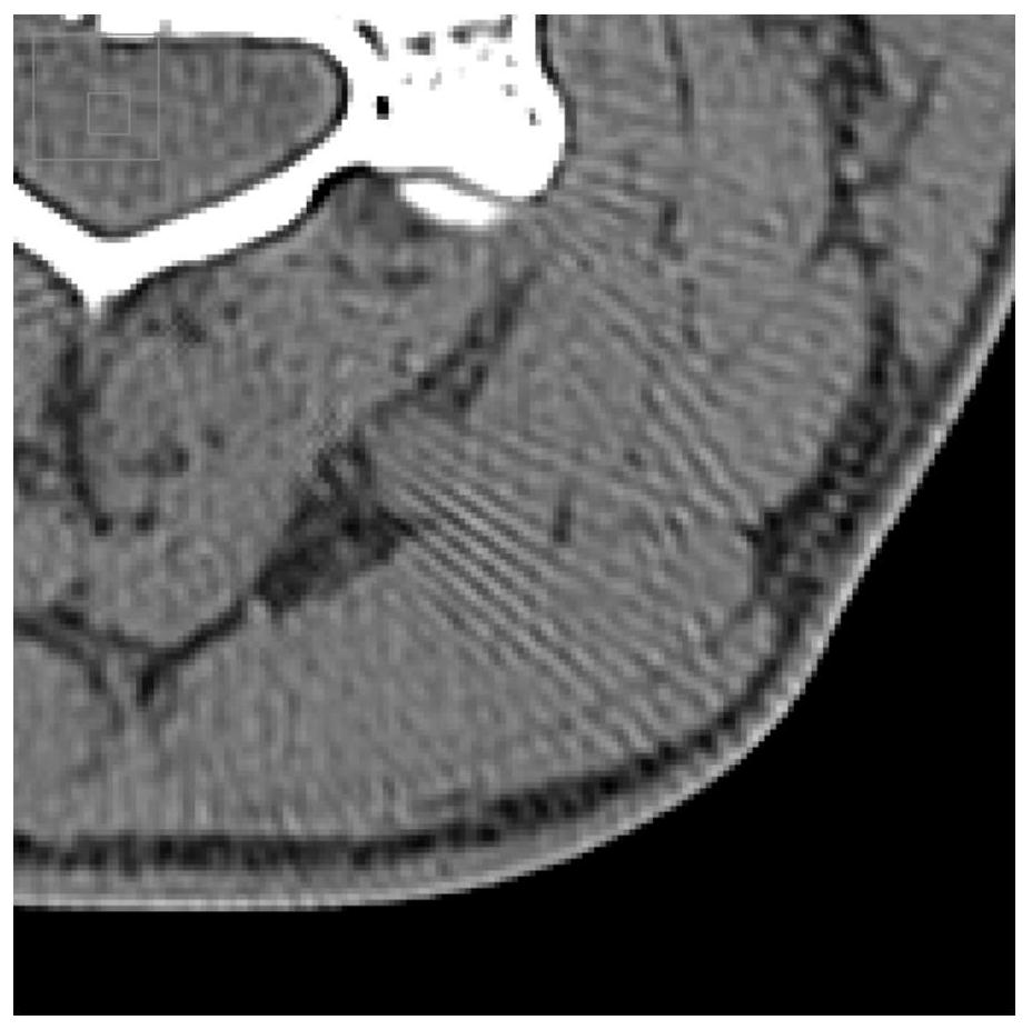
[0037] (1) input image; input image (marked as img in ,See figure 2 ).

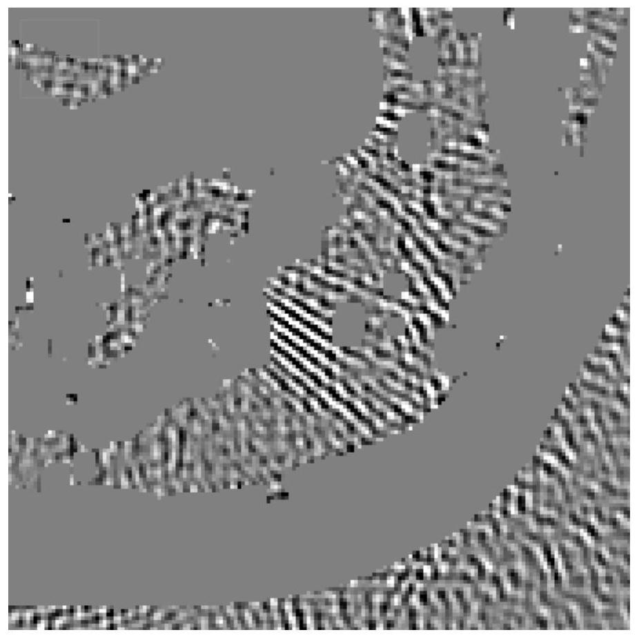
[0038] (2) Obtain an image texture with edge protection through median filtering and Laplacian filtering; specifically, median filtering and Laplacian filtering are respectively performed on the input image. The image texture distribution can be obtained by subtracting the input image from the result of median filtering. The edge distribution of the image can be obtained by the input image through Laplacian filtering. According to the result of Laplacian filtering, the s...


PUM


Abstract

The invention provides a CT image artifact removing method, which relates to the technical field of medical imagines.The method comprises the steps of filtering an input image to obtain an image texture with an edge protection area, obtaining phase distribution and amplitude distribution of the image texture at a frequency threshold, calculating and normalizing the mean value of the same frequency components in the amplitude distribution, obtaining normalized amplitude distribution, and obtaining peak distribution according to a set threshold value and a low-frequency mask; performing Gaussian filtering on the peak distribution to obtain a filtering matrix, and obtaining a non-aliasing texture frequency domain image; processing the non-aliasing texture frequency domain image according to the phase distribution of the image texture at the frequency threshold to obtain a non-aliasing texture space domain image; subtracting the image texture from the non-aliasing texture spatial domain image to obtain an aliasing spatial domain image; and finally, subtracting the aliasing artifact image from the input image to obtain an image after aliasing artifact elimination. According to the method, more image details can be reserved while aliasing artifacts are removed.

Description

technical field [0001] The invention relates to a method for correcting CT artifacts, in particular to a method for removing CT image artifacts, which belongs to the technical field of medical imaging. Background technique [0002] Aliasing artifact is a very common artifact in CT image reconstruction. This artifact is generally caused by insufficient frame sampling or insufficient detector sampling. Its appearance on the image is that radial stripes appear on the edge of the image or the edge of high-density materials. The existence of aliasing artifacts can reduce the image quality in mild cases, and affect clinical diagnosis in severe cases. [0003] In order to solve CT aliasing artifacts caused by insufficient frame sampling, a common method is to increase the sampling rate in the projection direction. In order to solve the aliasing artifacts caused by insufficient detector sampling, there are several common methods on the system hardware as follows: one is to use 1 / ...

Claims


Application Information

Patent Timeline
Patent Type & Authority Applications(China)
IPC IPC(8): G06T5/00G06T5/50
CPCG06T5/50G06T2207/10081G06T2207/20032G06T5/80G06T5/70
Inventor 徐亦飞陈伟潘华素
Owner FMI MEDICAL SYST CO LTD
Learn More