Promoter molecule capable of specifically recognizing proliferative endothelial cells and engineering cells
A technique for activating molecules and endothelial cells, applied in the field of bioengineering, can solve the problems of identifying new blood vessels that have not been reported, and achieve the effect of improving specificity
- Summary
- Abstract
- Description
- Claims
- Application Information
AI Technical Summary
Problems solved by technology
Method used
Image
Examples
Embodiment 1
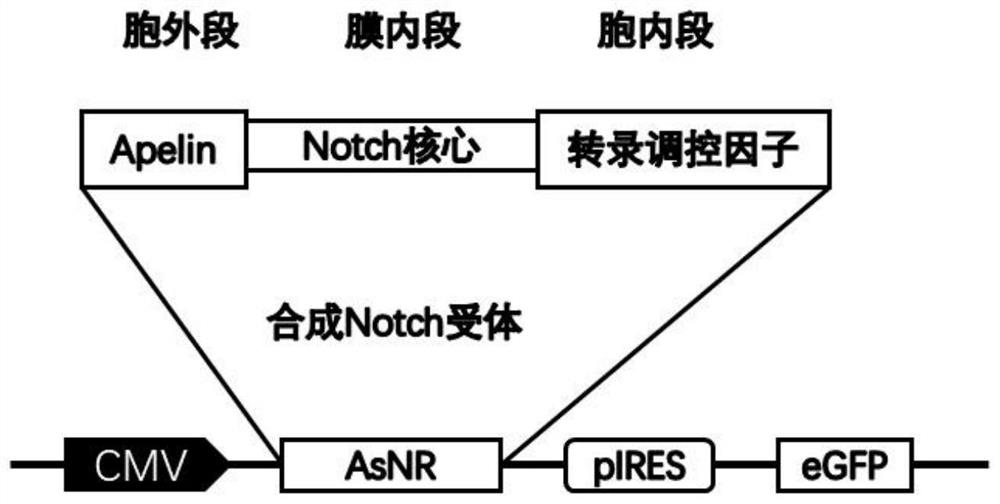
[0059] Embodiment 1 A preparation method of synNotch engineered endothelial progenitor cells, comprising the following steps:
[0060] 1) Preparation of endothelial progenitor cells
[0061] Take a C57 / BL6J mouse with a pregnancy period of 14-18 days, put the pregnant uterus in pre-cooled PBS containing 500-1000U of penicillin and streptomycin, soak for 3-5min, take out the fetal mouse, wash it twice with PBS; take it out under a microscope Fetal lung tissue was placed in PBS and shredded into small cubes with a diameter of about 1 mm.
[0062] Add 0.25% trypsin to the fetal lung tissue, culture in DMEM medium with 10% FBS for 16h-24h, centrifuge at 1500rpm for 5 minutes, discard the supernatant, take its precipitate, and use 5ml of EBM containing growth factor and 5%FBS serum- 2 medium to suspend cells to 10 6 / ml cells inoculated on T25cm 2 Cell culture flasks were cultured in a constant temperature incubator with 5% carbon dioxide at 37°C. When the growth and confluence...
Embodiment 2
[0078] Example 2 Co-cultivation of engineered cells and Apj-positive endothelial cells
[0079] 1. Isolation and culture of endothelial cells
[0080] Take the fresh umbilical cord of the postpartum mouse, cut off a 0.5 cm long section with surgical scissors; inject PBS solution into the umbilical vein, wash the residual blood; fill the umbilical vein with 0.1% collagenase, and digest it at room temperature for 5 minutes; Transfer the digestion solution containing endothelial cells to a 15ml centrifuge tube, centrifuge at 1000rpm for 5 minutes, discard the supernatant, add enough 1640 culture medium to resuspend, inoculate into a T25 culture bottle and culture for 2-3 days, the cells grow to 80- Passage at 90% confluence to obtain endothelial cells.
[0081] 2. Transfection and co-culture
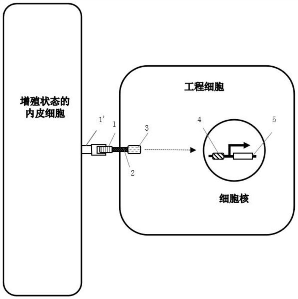
[0082] Transfect the CMV promoter molecule, TRE and Sphk1-Mfsd2a gene into the prepared endothelial progenitor cells. When the promoter molecule binds to Apj, tTA will detach from the cel...
Embodiment 3
[0085] Example 3 Engineered cells identify neovascularization after traumatic brain injury
[0086] (1) Construction of traumatic brain injury model
[0087] The mice were anesthetized by intraperitoneal injection of pentobarbital sodium (0.4mg / 10g), fixed on the stereotaxic injection apparatus, and then a hole with a diameter of about 5 mm was opened in the right parietal cortex with the central suture The lines are adjacent. Then a transducing metal rod with a diameter of 2.5 mm was placed on the skull opening, and a 5-gram gravity hammer was released from a height of 15 cm to make it fall freely and hit the transducing metal rod vertically.
[0088] (2) Simultaneous transfection of promoter molecules, transcriptional response element TRE, and red fluorescent protein RFP into endothelial progenitor cells;
[0089] (3) On the 3rd day after the TBI mouse model was successfully established, the engineered cells transformed from endothelial progenitor cells were injected into ...
PUM
 Abstract
Description
Claims
Application Information
 - R&D Engineer
- R&D Manager
- IP Professional
- Industry Leading Data Capabilities
- Powerful AI technology
- Patent DNA Extraction
Browse by: Latest US Patents, China's latest patents, Technical Efficacy Thesaurus, Application Domain, Technology Topic, Popular Technical Reports.
© 2024 PatSnap. All rights reserved.Legal|Privacy policy|Modern Slavery Act Transparency Statement|Sitemap|About US| Contact US: help@patsnap.com


