Premature infant retina plus lesion detection method and system

A premature infant retina and detection method technology, applied in the detection field of premature infant retinal plus lesions, can solve the problems of inability to compare and measure the retinal state, and achieve the effect of preventing disease delay

Pending Publication Date: 2021-03-09
智程工场(佛山)科技有限公司
View PDF0 Cites 1 Cited by
  • Summary
  • Abstract
  • Description
  • Claims
  • Application Information

AI Technical Summary

Problems solved by technology

[0003] Therefore, the technical problem to be solved by the present invention is to overcome the defect in the prior art that the detection results of retinal plus lesions in premature infants cannot objectively compare and measure the state of the retina, thereby providing a detection method and system for retinal plus lesions in premature infants , to quantify the degree of lesion by converting retinal images of premature infants into corresponding i-ROP lesion scores, which can objectively and accurately measure the current lesion status

Method used


Image

  • Premature infant retina plus lesion detection method and system
  • Premature infant retina plus lesion detection method and system
  • Premature infant retina plus lesion detection method and system

Examples


Embodiment 1

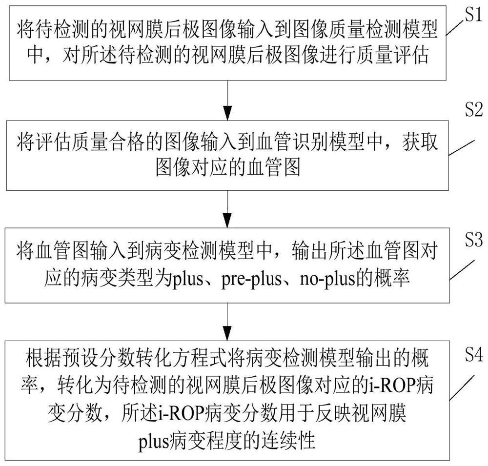
[0037] Embodiments of the present invention provide a method for detecting retinal plus lesions in premature infants, such as figure 1 As shown, the method includes the following steps:

[0038] Step S1: Input the image of the retinal posterior pole to be detected into the image quality detection model, and evaluate the quality of the image of the retinal posterior pole to be detected.

[0039] In the embodiment of the present invention, it is evaluated whether the retinal posterior pole image to be detected satisfies the standards of retinal image, retinal posterior pole image and high-quality image at the same time. In order to make a correct diagnosis of the severity of ROP, if the quality of the image is not up to standard, the image will not undergo the subsequent inspection process, and the quality assurance of the image can be realized by evaluating the quality of the image. In a specific embodiment, the image quality detection model is a neural network model trained f...

Embodiment 2

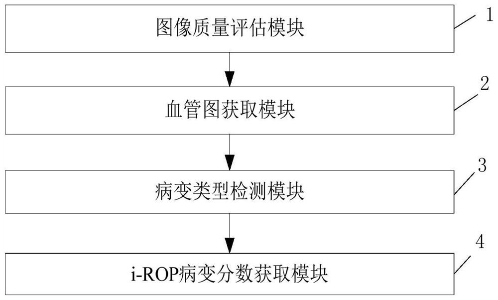
[0054] Embodiments of the present invention provide a detection system for retinal plus lesions in premature infants, such as figure 2 shown, including:

[0055] Image quality evaluation module 1, for inputting the retinal posterior pole image to be detected into the image quality detection model, and performing quality assessment on the retinal posterior pole image to be detected; this module executes the description of step S1 in embodiment 1 method, which will not be repeated here.

[0056] The blood vessel map acquisition module 2 is used to input the image with qualified evaluation quality into the blood vessel recognition model, and obtain the blood vessel map corresponding to the image; this module executes the method described in step S2 in Embodiment 1, which will not be repeated here.

[0057] The lesion type detection module 3 is used to input the blood vessel map into the lesion detection model, and output the probability that the lesion type corresponding to the...

Embodiment 3

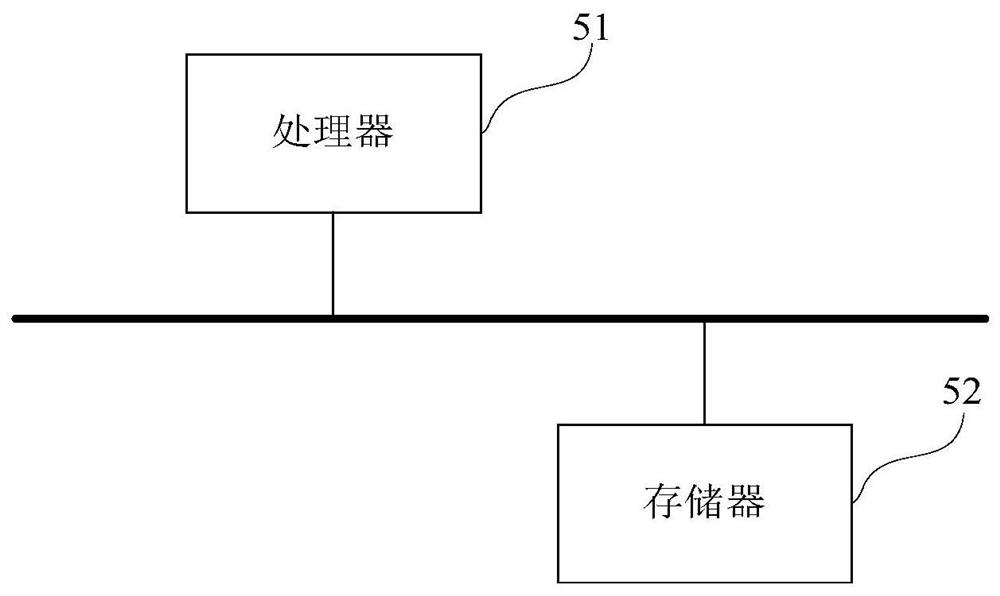
[0061] An embodiment of the present invention provides a computer device, such as image 3 As shown, the device may include a processor 51 and a memory 52, wherein the processor 51 and the memory 52 may be connected via a bus or in other ways, image 3 Take connection via bus as an example.

[0062] The processor 51 can be a central processing unit (Central Processing Unit, CPU), and can also be other general processors, digital signal processors (Digital Signal Processor, DSP), application specific integrated circuits (Application Specific Integrated Circuit, ASIC), on-site Programmable gate array (Field-Programmable Gate Array, FPGA) or other programmable logic devices, discrete gate or transistor logic devices, discrete hardware components and other chips, or a combination of the above-mentioned types of chips.


PUM


Abstract

The invention discloses a premature infant retina plus lesion detection method and system, and the method comprises the steps: firstly inputting a to-be-detected retina posterior pole image into an image quality detection model for quality evaluation, and inputting a qualified image into a blood vessel recognition model to obtain a blood vessel graph; inputting the blood vessel graph into a lesiondetection model, and outputting the probability of the corresponding lesion types of plus, pre-plus and no-plus; and converting the probability of the lesion type of the lesion examination into a corresponding i-ROP lesion score according to a preset score conversion equation to reflect the continuity of the retinopathy degree. According to the invention, the severity of plus lesion is objectively measured, and the severity is tracked over time to provide objective assessment of disease progression or regression; by analyzing the change of the score along with the time, the eyes which progress into plus lesion ROP can be recognized in advance under most conditions for early intervention, disease delay caused by insufficient diagnosis is prevented, the accuracy of lesion degree detection is high, and the ophthalmoscope examination frequency of doctors can be greatly reduced.

Description

technical field [0001] The invention relates to the technical field of image recognition, in particular to a method and system for detecting retinal plus lesions in premature infants. Background technique [0002] Plus lesions are the most important clinical criteria to determine the need for ROP (retinopathy of prematurity) treatment, and failure to correctly diagnose plus lesions increases the risk of irreversible blindness. According to the International Classification of ROP (ICROP), plus lesions are divided into 3 grades (no plus, pre-plus or plus lesions), but the diagnostic inconsistencies between observers due to subjective factors lead to differences in diagnostic results. The existing ROP automatic diagnosis methods all use the diagnostic model to automatically diagnose which of the three grades the lesion is in, but they cannot give precise guidance on the degree of the lesion, and thus cannot objectively compare and measure the degree of retinopathy in premature ...

Claims


Application Information

Patent Timeline
IPC IPC(8): G06T7/00
CPCG06T7/0012G06T2207/20081G06T2207/20084G06T2207/30041G06T2207/30096G06T2207/30168
Inventor 彭晟
Owner 智程工场(佛山)科技有限公司
Why Eureka
Try Eureka