Automatic analysis system and method for full-slice digital pathological image

An automatic analysis system and digital pathology technology, applied in the field of medical image processing, can solve problems such as high requirements for doctors' professional ability, high requirements for researchers' professional knowledge, and long time consumption, so as to improve the efficiency of diagnosis and improve the accuracy of diagnosis. The effect of accuracy

Pending Publication Date: 2021-03-02
SHANDONG PROVINCIAL HOSPITAL AFFILIATED TO SHANDONG FIRST MEDICAL UNIVERSITY
View PDF6 Cites 20 Cited by
  • Summary
  • Abstract
  • Description
  • Claims
  • Application Information

AI Technical Summary

Problems solved by technology

[0004] The inventors found that the following problems still exist in the analysis of pathological images: the doctor’s diagnosis takes a long time, and the doctor’s professional ability is very high; the traditional machine learning method for analyzing pathological images mainly depends on the effect of feature extraction , which requires high professional knowledge of researchers; the existing deep learning-based methods can only analyze small-sized images, but the size of digital pathology images is too large, and the existing methods cannot be directly used for large-sized full-slice digital images. Analysis of pathological images

Method used


Image

  • Automatic analysis system and method for full-slice digital pathological image
  • Automatic analysis system and method for full-slice digital pathological image
  • Automatic analysis system and method for full-slice digital pathological image

Examples


Embodiment 1

[0038]The purpose of this embodiment is to provide an automatic analysis system for full-slice digital pathological images.

[0039]An automatic analysis system for full-slice digital pathological images, including:

[0040]Image preprocessing module: it is used to divide the acquired full-slice digital pathology image into several image pieces; divide the pathology image pieces into background blank area and foreground tissue area to obtain image pieces of foreground tissue area; according to training set samples Color distribution performs color conversion on the small image blocks to enhance the color variability of the data;

[0041]Wherein, the image blocks are named according to a certain naming rule, and are used for image mosaic restoration after processing.

[0042]Automatic analysis module: it is used to analyze and process the processed image small blocks using the pre-trained deep learning model, and realize the analysis of the full-slice pathological image according to the processi...

Embodiment 2

[0097]The purpose of this embodiment is to provide an automatic analysis method for full-slice digital pathological images.

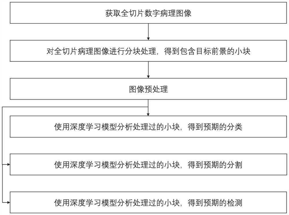
[0098]An automatic analysis method for full-slice digital pathological images, including:

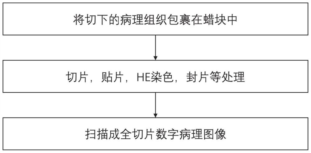
[0099]Divide the acquired full-slice digital pathology image into several image pieces;

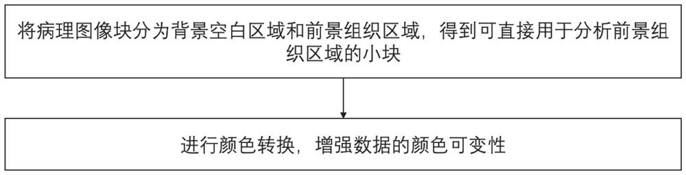
[0100]Divide the pathological image into the background blank area and the foreground tissue area, and obtain the image of the foreground tissue area;

[0101]Performing color conversion on the small image blocks according to the color distribution of the training set samples to enhance the color variability of the data;

[0102]The pre-trained deep learning model is used to analyze and process the processed image small blocks, and the analysis of the full-slice digital pathological image is realized according to the analysis and processing result.

[0103]The present disclosure provides an automatic analysis method and system for full-slice digital histopathological images, using a deep learning m...

Embodiment 3

[0105]The purpose of this embodiment is to provide an electronic device.

[0106]An electronic device includes a memory, a processor, and a computer program stored and running on the memory, and the processor implements the automatic analysis method of a full-slice digital pathological image when the processor executes the program, and includes:

[0107]Divide the acquired full-slice digital pathology image into several image pieces;

[0108]Divide the pathological image into the background blank area and the foreground tissue area, and obtain the image of the foreground tissue area;

[0109]Performing color conversion on the small image blocks according to the color distribution of the training set samples to enhance the color variability of the data;

[0110]The pre-trained deep learning model is used to analyze and process the processed image small blocks, and the analysis of the full-slice digital pathological image is realized according to the analysis and processing result.


PUM


Abstract

The invention provides an automatic analysis system and method for a full-slice digital pathological image. The method comprises the steps of: obtaining the full-slice digital pathological image; carrying out partitioning processing on the full-slice digital pathological image; dividing the pathological image block into a background blank area and a foreground tissue area, and executing color normalization on the foreground area image block to obtain small blocks which can be directly used for analyzing the foreground tissue area; after image preprocessing, classifying the pathological image blocks to obtain a predictor type classification result, a segmentation result and a target detection result of each pathological image small block; after a full-slice digital pathological image is acquired, partitioning and preprocessing the full-slice digital pathological image, so that image classification, segmentation and target detection tasks can be carried out by using a deep learning model, and then visual display is carried out, thereby assisting a pathologist to observe and analyze the image, and improving the diagnosis efficiency and the diagnosis accuracy.

Description

Technical field[0001]The present disclosure relates to the technical field of medical image processing, and in particular to an automatic analysis system and method for full-slice digital pathological images.Background technique[0002]Histopathological examination is a pathological method used to examine the pathological changes in tissues to analyze and diagnose diseases. It is currently the most accurate method for diagnosing cancer. Pathological images can help doctors analyze the patient's condition and obtain the specific conditions of tumor cells, such as the degree of differentiation, whether there is lymph node metastasis, etc., which are helpful in diagnosis, staging, and prognosis of the disease. Under normal circumstances, pathologists classify and type the tissues and obtain a histopathology report to determine the next treatment plan. Using scanning imaging equipment to take pathological specimens, high-quality digital images of pathology can be obtained.[0003]In recent ...

Claims


Application Information

Patent Timeline
Patent Type & Authority Applications(China)
IPC IPC(8): G06T7/00G06K9/62G06T7/10G16H30/00G16H50/20
CPCG06T7/0012G06T7/10G16H50/20G16H30/00G06T2207/30004G06F18/241G06F18/214
Inventor 肖伟郑元杰姚志刚姜岩芸周小明隋晓丹高远马帅
Owner SHANDONG PROVINCIAL HOSPITAL AFFILIATED TO SHANDONG FIRST MEDICAL UNIVERSITY
Why Eureka
Try Eureka