Tissue blood oxygen imaging detection method based on two-stage spatial mapping

A space mapping and imaging detection technology, applied in image enhancement, image analysis, image data processing, etc., can solve the problem of universality and accuracy that cannot meet the requirements of clinical applications, reduce the real-time accuracy of detection results, and limit application scenarios, etc. problems, to achieve the effect of simplifying the process of tissue blood oxygen parameters, improving the accuracy of estimation, and ensuring the speed of calculation

Active Publication Date: 2021-01-05
SHANGHAI JIAO TONG UNIV +1
View PDF10 Cites 3 Cited by
  • Summary
  • Abstract
  • Description
  • Claims
  • Application Information

AI Technical Summary

Problems solved by technology

These methods have strong restrictions on application scenarios, and their universality and accuracy cannot meet the requirements of actual clinical applications.
The main reason is that these methods equate the tissue in the surgical field to an ideal tissue with a single composition (oxygenated and deoxygenated hemoglobin), and the anatomical structure information and other components of the tissue are ignored; in addition, Lambert-Beer's law is directly used to construct The equations, which are equivalent to the analysis and solution of transmission imaging, do not take into account the characteristics of the photons detected in the actual reflection imaging through multiple scattering and diffusion of the tissue, which will undoubtedly reduce the real-time accuracy of the detection results

Method used


Image

  • Tissue blood oxygen imaging detection method based on two-stage spatial mapping
  • Tissue blood oxygen imaging detection method based on two-stage spatial mapping
  • Tissue blood oxygen imaging detection method based on two-stage spatial mapping

Examples


Embodiment

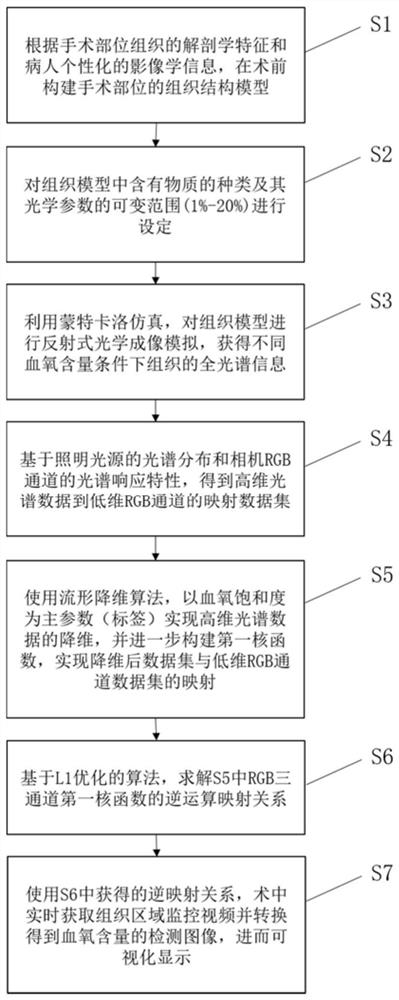
[0055] Such as figure 1 As shown, a tissue oxygen imaging detection method based on two-stage spatial mapping includes the following steps:

[0056] S1. Construct a tissue model according to the anatomical characteristics of the tissue and individual imaging data, and determine the tissue structure parameters. If there is no individual imaging data, the variable range of the tissue structure parameters needs to be set;

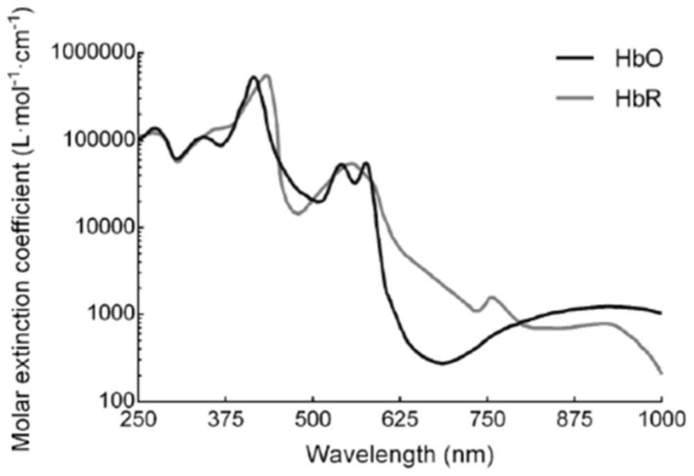
[0057] S2. Determine the types of substances contained in the tissue, and set the variable range of optical parameters of each type of substance, wherein the optical parameters include absorption coefficient, scattering coefficient, refractive index and anisotropy parameters;

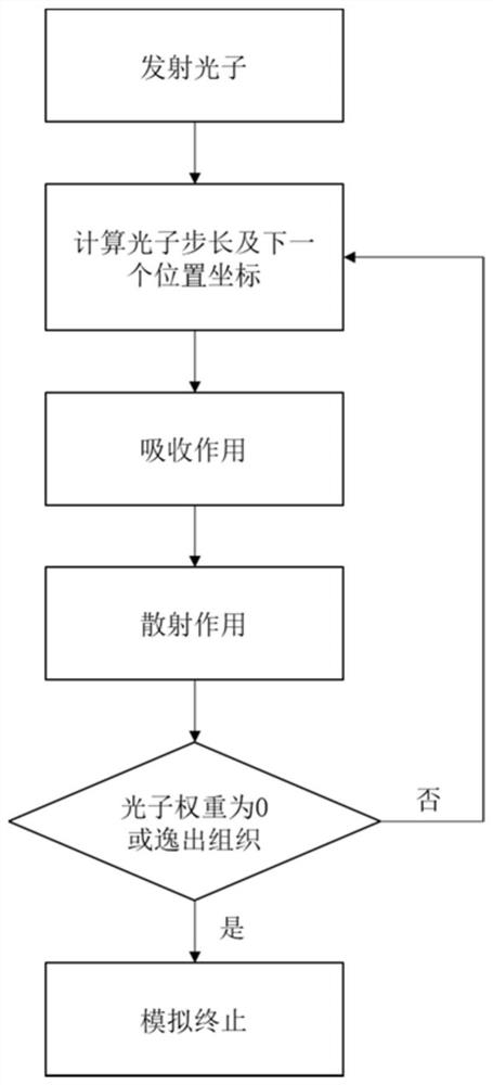
[0058] S3. Through Monte Carlo simulation, reflective optical imaging simulation is performed on the tissue model to obtain tissue structure parameters and optical parameters of various material types corresponding to various combinations within the variable range under different blood ...


PUM


Abstract

The invention relates to a tissue blood oxygen imaging detection method based on two-stage spatial mapping. The tissue blood oxygen imaging detection method comprises the following steps of: constructing a tissue structure model before an operation; setting material types and optical parameters in tissues; performing reflection type optical imaging simulation to obtain high-dimensional full-spectrum information; obtaining a mapping data set of a low-dimensional RGB channel based on the spectral characteristics of an illumination light source and an imaging camera; by using a manifold dimensionality reduction algorithm, achieving dimensionality reduction of high-dimensional spectral data with the blood oxygen saturation as a main parameter, constructing a first kernel function, and achieving mapping of a data set obtained after dimensionality reduction and a low-dimensional RGB channel data set; obtaining an inverse operation mapping relation of the first kernel function based on an L1optimization strategy; and, imaging the actual to-be-detected tissue in the operation, and calculating in real time through the adjusted inverse operation mapping relation to obtain an estimated valueof the blood oxygen saturation. Compared with the prior art, the two-stage spatial mapping relation is utilized in the invention; the accuracy of blood oxygen parameter estimation can be effectivelyimproved; and meanwhile, the real-time performance of imaging detection is guaranteed.

Description

technical field [0001] The present invention relates to the technical field of imaging detection, in particular to a tissue blood oxygen imaging detection method based on two-stage spatial mapping, so as to realize real-time and accurate detection of intraoperative tissue blood oxygen directly by using intraoperative white light visual field RGB images, and its purpose is not to Obtain a diagnosis, state of health or treatment. Background technique [0002] A large number of clinical operations have shown the importance of real-time blood oxygen information detection, especially in the field of surgery, real-time monitoring of blood oxygen distribution and changes can effectively reduce surgical risks and improve surgical outcomes. In order to realize the detection of physiological parameters such as (de)oxyhemoglobin content, blood volume, and blood oxygen saturation, tissue perfusion is often used at present. Tissue perfusion is a fluorescence imaging method with the help ...

Claims


Application Information

Patent Timeline
Patent Type & Authority Applications(China)
IPC IPC(8): A61B5/1455G06T7/00G06K9/62
CPCA61B5/14551A61B5/14542G06T7/0012G06T2207/10024G06T2207/30101G06F18/21375
Inventor 童善保禹婧祎苗鹏
Owner SHANGHAI JIAO TONG UNIV
Why Eureka
Try Eureka