An endoscopic oct-raman dual-mode imaging device and imaging method

A dual-mode imaging and imaging technology, which is applied in the field of endoscopy, can solve the problems of affecting Raman signal strength, uniform image distortion, and uneven speed, so as to improve the degree of visualization, solve image distortion, and ensure accuracy.

Active Publication Date: 2022-04-05
南京影衍医疗科技有限公司
View PDF19 Cites 0 Cited by
  • Summary
  • Abstract
  • Description
  • Claims
  • Application Information

AI Technical Summary

Problems solved by technology

[0004] The existing OCT-Raman endoscopic probe has the following problems: First, although the existing Raman endoscopic system can obtain the Raman spectrum of the endoscopic image and its corresponding position, the Raman spectrum itself exists in the form of spectral lines , complex data analysis is required to realize the analysis of lesions, the degree of correlation with the imaging endoscopic image is low, the position correspondence is not obvious, the degree of visualization is low, and the abnormality and lesion cannot be clearly judged
Secondly, in the cavity endoscopy system, the imaging position of the endoscopic probe is limited by the position of the imaging catheter in the cavity, and its imaging center is not in the center of the blood vessel. Although this has no great impact on endoscopic imaging, it is very important for Raman detection The detection distance greatly affects the intensity of the Raman signal
Finally, the mechanical rotation scanning of most endoscopic catheters is realized by torsion cables. In the detection of large-length cavities, there is a problem of uneven rotation speed that leads to uniform image distortion, and its rotation speed and stability are affected to a certain extent. degree of limitation

Method used


Image

  • An endoscopic oct-raman dual-mode imaging device and imaging method
  • An endoscopic oct-raman dual-mode imaging device and imaging method
  • An endoscopic oct-raman dual-mode imaging device and imaging method

Examples


Embodiment Construction

[0034] In order to make the object, technical solution and advantages of the present invention clearer, the present invention will be further described in detail below in conjunction with the accompanying drawings and embodiments. It should be understood that the specific embodiments described here are only used to explain the present invention, not to limit the present invention.

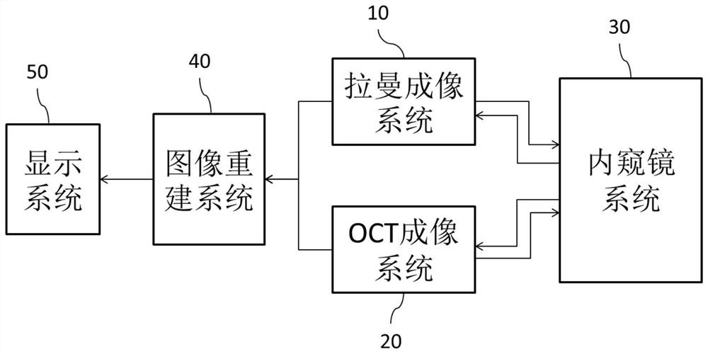
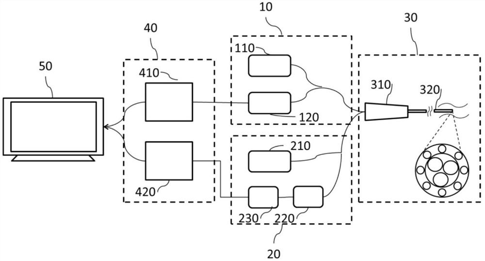
[0035] figure 1 It is a schematic diagram of the overall structure of the endoscopic OCT-Raman dual-mode imaging device of the embodiment of the present invention, figure 2It is a connection schematic diagram of the endoscopic OCT-Raman dual-mode imaging device of the embodiment of the present invention. In this embodiment, the endoscopic OCT-Raman dual-mode imaging device includes a Raman imaging system 10 , an OCT imaging system 20 , an endoscope system 30 , an image reconstruction system 40 and a display system 50 . Endoscope system 30 is connected with Raman imaging system 10 and OCT imaging...


PUM


Abstract

The invention discloses an endoscopic OCT-Raman dual-mode imaging device and an imaging method, including a Raman imaging system, an OCT imaging system, an endoscope system, an image reconstruction system and a display system. The endoscope system is respectively connected with the Raman imaging system and the OCT imaging system, both of the Raman imaging system and the OCT imaging system are connected with the image reconstruction system, and the image reconstruction system is connected with the display system. The invention can obtain a three-dimensional image with higher optical resolution, changes the existence mode of the spectral line form of the Raman spectrum, associates the Raman spectrum with the OCT three-dimensional imaging, and realizes the tiled three-dimensional image of the OCT cavity and its corresponding Raman spectrum. The reconstruction of the MAN 3D image makes the correspondence of the lesion position more obvious, improves the visualization of the imaging, and realizes the accurate and rapid acquisition of OCT imaging data and Raman signals.

Description

technical field [0001] The invention belongs to the technical field of endoscopes, in particular to an endoscope OCT-Raman dual-mode imaging device and an imaging method. Background technique [0002] As a non-invasive or minimally invasive imaging method, endoscopy can penetrate deep into the body cavity of the living body and has been widely used in the imaging diagnosis of multiple cavities such as blood vessels, digestive tract, urinary system and respiratory tract. OCT is a high-resolution imaging and observation method of mesoscopic (micron-scale) living tissue using the coherence principle of scattered light from biological tissue. OCT endoscope is an important branch of endoscopic technology. While OCT technology has high resolution, it also has large tomographic capabilities, and can be easily combined with fiber optic catheters and endoscopes to image internal organs of the human body. In particular, endoscopic frequency-domain OCT can realize high-speed real-time...

Claims


Application Information

Patent Timeline
Patent Type & Authority Patents(China)
IPC IPC(8): A61B5/00A61B1/07
CPCA61B5/0066A61B5/0075A61B5/7203A61B5/7225A61B1/07A61B1/0646
Inventor 陈春晓徐俊琪陈志颖
Owner 南京影衍医疗科技有限公司
Why Eureka
Try Eureka