
Leukemia cell microscopic image classification method and system based on DenseNet

A leukemia cell and microscopic image technology, which is applied in the field of leukemia cell microscopic image classification method and system, can solve problems such as difficult to distinguish, pixel intensity value is not an ideal distinguishing feature, and difficult to identify, so as to reduce the missed diagnosis rate and misdiagnosis rate , Auxiliary diagnosis of leukemia, and the effect of improving classification ability

Active Publication Date: 2020-12-15
XIAMEN UNIV
View PDF4 Cites 2 Cited by
  • Summary
  • Abstract
  • Description
  • Claims
  • Application Information

AI Technical Summary

Problems solved by technology

However, because the morphology of leukemia cells and normal cells is very similar, it is difficult to distinguish, the traditional manual diagnosis method is time-consuming and laborious, and is greatly affected by the doctor's subjective, there are problems of missed diagnosis and misdiagnosis
[0003] However, using deep learning algorithms to automatically classify leukemia cell microscopic images is also facing difficulties, mainly because: in general, when convolutional neural networks deal with image classification, detection and other recognition problems, they are all performed in the RGB color space Yes, but because the positive class (leukemic cells) and the negative class (normal cells) in the leukemia cell microscopic image are very similar in morphology, the intensity value of the pixel in the RGB space is not an ideal distinguishing feature. In the RGB color space It is more difficult to identify under the environment, and the effect is poor

Method used


Image

  • Leukemia cell microscopic image classification method and system based on DenseNet
  • Leukemia cell microscopic image classification method and system based on DenseNet
  • Leukemia cell microscopic image classification method and system based on DenseNet

Examples


Embodiment 1

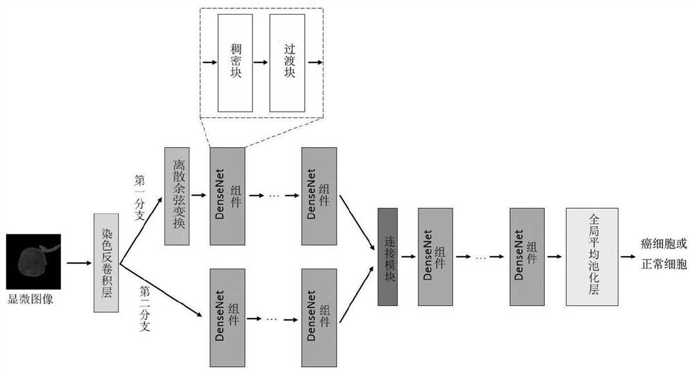
[0033] A DenseNet-based classification method for leukemia cell microscopic images, such as figure 1 shown, including the following steps:

[0034] S1. Input a microscopic image of leukemia cells, input it into the dyeing deconvolution layer for processing, and calculate the dye absorption of the cells;

[0035] S2, the first branch processes the dye absorption amount through discrete cosine transform, extracts the frequency domain information, and then transmits to the subsequent DenseNet component for processing to obtain the feature map in the frequency threshold;

[0036] S3. The second branch directly transfers the dye absorption amount outputted in step S1 to the DenseNet component for processing to obtain a feature map in the spatial domain;

[0037] S4. Connect the two feature maps output by the first branch and the second branch in the channel direction, fuse the optical density space dye absorption feature and its feature information in the frequency domain, and the...


PUM


Abstract

The invention discloses a leukemia cell microscopic image classification method and system based on DenseNet, and the method comprises the steps: adding a dyeing deconvolution layer and a discrete cosine transform module on the basis of a DenseNet network, so as to obtain the feature information of a cell microscopic image in a spatial domain and a frequency threshold of coloring agent absorptionamount; and acquiring a feature vector by fusing the two kinds of information and using the vector for judging whether the blood cells belong to cancer cells or not. The coloring agent absorption amount is used for representing the characteristics of the cell microscopic image, the problem that recognition is difficult in an RGB color space is solved, the average accuracy rate and the average weighted F1 score reach 89.81% and 89.64% respectively, automatic classification of leukemia cells is achieved, and therefore a pathologist is assisted in diagnosing leukemia, and the missed diagnosis rate and the misdiagnosis rate are reduced.

Description

technical field [0001] The invention relates to the technical field of image processing, in particular to a method and system for classifying leukemia cell microscopic images based on DenseNet. Background technique [0002] Early diagnosis of leukemia is very important to improve the survival rate of patients. The traditional method of identifying leukemia cells is performed by a pathologist looking at the characteristics of the cells in a blood smear under a light microscope. However, because the morphology of leukemia cells is very similar to that of normal cells, it is difficult to distinguish. The traditional manual diagnosis method is time-consuming and laborious, and is greatly influenced by the doctor's subjectivity, so there are problems of missed diagnosis and misdiagnosis. [0003] However, using deep learning algorithms to automatically classify leukemia cell microscopic images is also facing difficulties, mainly because: in general, when convolutional neural net...

Claims


Application Information

Patent Timeline
Patent Type & Authority Applications(China)
IPC IPC(8): G06K9/00G06K9/46G06K9/62G06N3/04G06N3/08
CPCG06N3/08G06V20/695G06V20/698G06V10/56G06N3/045G06F18/253
Inventor 王连生
Owner XIAMEN UNIV
Learn More