Thyroid malignant nodule detection method based on deep learning

A detection method and deep learning technology, applied in the field of image processing, can solve problems such as heavy workload, and achieve the effects of reducing errors, good robustness, and high detection accuracy

Pending Publication Date: 2020-11-27
TIANJIN UNIV
View PDF5 Cites 1 Cited by
  • Summary
  • Abstract
  • Description
  • Claims
  • Application Information

AI Technical Summary

Problems solved by technology

However, because ultrasound diagnosis requires doctors to manually mark the lesion area, the workload is heavy, and doctors with different experience and levels often have subjective diagnosis of the results.

Method used


Image

  • Thyroid malignant nodule detection method based on deep learning
  • Thyroid malignant nodule detection method based on deep learning
  • Thyroid malignant nodule detection method based on deep learning

Examples


Embodiment Construction

[0040] The present invention will be further described in detail below through the specific examples, the following examples are only descriptive, not restrictive, and cannot limit the protection scope of the present invention with this.

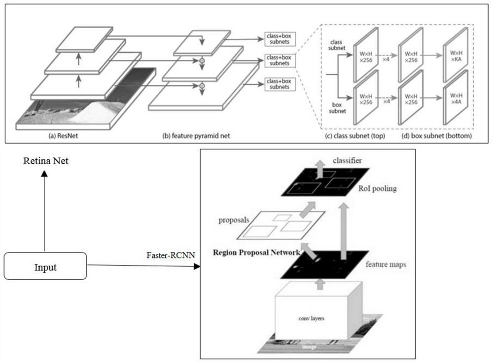
[0041] A method for detecting malignant thyroid nodules based on deep learning, characterized in that: the steps of the method are:

[0042] S0101: Input ultrasonic medical images for model training and detection, and the program will remove some additional marks contained in the image data, including the size of the nodule, the name and model of the device, and patient privacy;

[0043] S0102: Save the pictures into JPEGImages files and name them in a unified format;

[0044] S0103: Divide the data set, generate a .txt file under the Main folder, including the verification set, training set, and test set image number, use the labelImg tool to label the image, create a box border for the target object, and save it to generate an .xml file, i...


PUM


Abstract

The invention relates to a thyroid malignant nodule detection method based on deep learning, the method is used for the auxiliary diagnosis of thyroid malignant nodules, and the automatic labeling ofa region of interest can be achieved after the training of a large amount of data under the characteristics of low medical image resolution, low precision and low target and background identificationdegree. Errors caused by subjective factors can be effectively reduced, and radiologists can be helped to diagnose quickly and accurately. The detection precision and speed are considerable, and the method has the potential of clinical application.

Description

technical field [0001] The invention belongs to the field of image processing, and relates to deep learning technology and target detection technology, in particular to a method for detecting malignant thyroid nodules based on deep learning. Background technique [0002] Ultrasonic diagnostic technology is widely used in clinical practice due to its advantages of convenient inspection and low cost. Studies have shown that ultrasound has advantages in differentiating benign and malignant thyroid nodules, because there are obvious differences between benign and malignant thyroid nodules in ultrasound images in terms of size, shape, number, cystic change, calcification, and blood supply, so clinicians These characteristics can be used to judge the nature of nodules. However, because ultrasound diagnosis requires doctors to manually mark the lesion area, the workload is heavy, and doctors with different experience and levels often have subjective diagnosis of the results. Ther...

Claims


Application Information

Patent Timeline
Patent Type & Authority Applications(China)
IPC IPC(8): G06T7/00G16H30/20G06N3/04
CPCG06T7/0012G16H30/20G06T2207/20081G06T2207/10132G06T2207/30096G06N3/045
Inventor 李雪威曾晨于瑞国刘志强喻梅高洁徐天一查涛
Owner TIANJIN UNIV
Why Eureka
Try Eureka