Brain magnetic resonance image segmentation method based on improved fuzzy C-means
A magnetic resonance image, blurring technology, applied in the field of medical image processing, can solve the problems of complex objective function and fitness function, no consideration of pixel spatial information, and slow optimization speed
- Summary
- Abstract
- Description
- Claims
- Application Information
AI Technical Summary
Problems solved by technology
Method used
Image

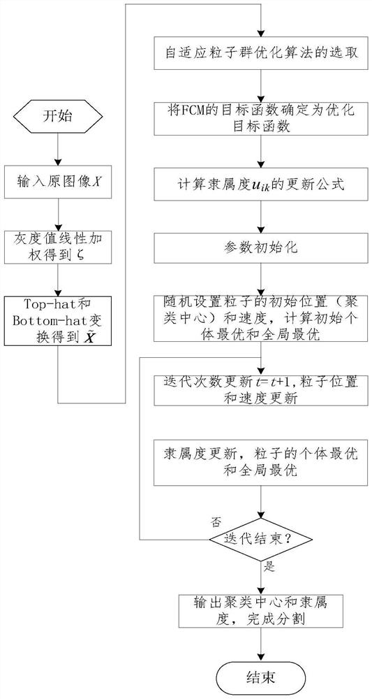

Examples
Embodiment 1
[0092] The test parameters of embodiment 1 and 2 (the value of these test parameters is the routine value in this area, can also obtain in conjunction with the research of relevant algorithm and a large amount of simulation experiments) settings are: population size (p=100), constant ( σ=40), ambiguity (m=2), maximum number of iterations (t max =500), error threshold (β=0.00001) and learning factor (c1=c2=2), in addition, for embodiment 1, division class number c=3, for embodiment 2, division class number c=4 .
[0093] 2. Simulation content:
[0094] Embodiment 1: adopt general FCM, GAFCM (genetic algorithm combined with fuzzy C mean value), FGFCM (fast generalized fuzzy C mean value), FRFCM (fast robustness fuzzy C mean value) and segmentation method of the present invention to image not containing cerebrospinal fluid respectively Carry out segmentation processing, the result is as follows figure 2 , where 2(a) is the source image containing noise, figure 2 (b1), 2(b2)...
Embodiment 2
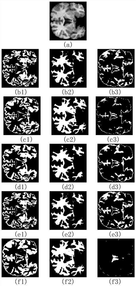
[0095] Embodiment 2: respectively adopt general FCM, GAFCM, FGFCM, FRFCM and the method of the present invention to carry out segmentation processing to the image containing cerebrospinal fluid, the result is as follows image 3 , where 3(a) is the source image containing noise, image 3 (b1), 3(b2) and 3(b3) are the gray matter, white matter and cerebrospinal fluid results obtained by the general FCM segmentation method, respectively, image 3 (c1), 3(c2) and 3(c3) are the gray matter, white matter and cerebrospinal fluid images obtained by GAFCM segmentation method respectively, image 3 (d1), 2(d2) and 3(d3) are the result images of gray matter, white matter and cerebrospinal fluid obtained by FGFCM segmentation method respectively, image 3 (e1), 3(e2) and 3(e3) are the result images of gray matter, white matter and cerebrospinal fluid obtained by FRFCM segmentation method respectively, image 3 (f1), 3(f2) and 3(f3) are gray matter, white matter and cerebrospinal fluid ...
PUM
 Abstract
Description
Claims
Application Information
 - R&D
- Intellectual Property
- Life Sciences
- Materials
- Tech Scout
- Unparalleled Data Quality
- Higher Quality Content
- 60% Fewer Hallucinations
Browse by: Latest US Patents, China's latest patents, Technical Efficacy Thesaurus, Application Domain, Technology Topic, Popular Technical Reports.
© 2025 PatSnap. All rights reserved.Legal|Privacy policy|Modern Slavery Act Transparency Statement|Sitemap|About US| Contact US: help@patsnap.com