Method and device for establishing cervical spinal cord simulation model
A simulation model and establishment method technology, applied in medical simulation, instruments, medical informatics, etc., can solve the problems of incomplete model anatomy, low verification and simulation, and achieve strong clinical significance, high simulation, Anatomically intact effect
- Summary
- Abstract
- Description
- Claims
- Application Information
AI Technical Summary
Problems solved by technology
Method used
Image
Examples
Embodiment 1
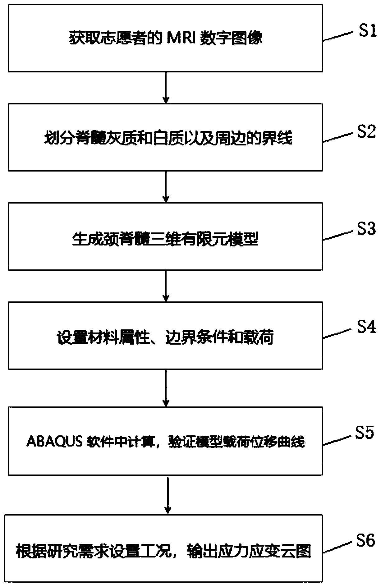
[0040] See figure 1 , figure 1 It is a flow chart of a method for establishing a cervical spinal cord simulation model disclosed in Embodiment 1. The method comprises the steps of:
[0041] Step S1: Acquire the MRI digital images of the cervical spinal cord of the volunteers, and save them as data files in DICOM format.
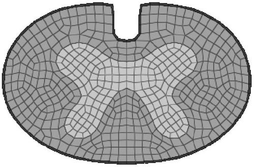
[0042] Step S2: Import the data in DICOM format into the medical imaging processing software Simpleware, and divide the gray matter and white matter of the cervical spinal cord and the boundary between the white matter and the surrounding areas according to the anatomical structure of the gray matter and white matter of the spinal cord and the difference in gray level in the image, and generate a cervical spinal cord transection surface model, and export boundary curves as IGES format files.
[0043] Step S3: Import the IGES format file into the finite element modeling software Hypermesh to generate a cross-sectional geometric model of the cervical spina...
Embodiment 2
[0053] See Figure 4 , Figure 4 It is a flow chart of another method for establishing a cervical spinal cord simulation model disclosed in Embodiment 2. The method comprises the steps of:
[0054] Step S1: Acquire the MRI digital images of the cervical spinal cord of the volunteers, and save them as data files in DICOM format.
[0055] Step S2: Import the data in DICOM format into the medical imaging processing software Simpleware, and divide the gray matter and white matter of the cervical spinal cord and the boundary between the white matter and the surrounding areas according to the anatomical structure of the gray matter and white matter of the spinal cord and the difference in gray level in the image, and generate a cervical spinal cord transection surface model, and export boundary curves as IGES format files.
[0056] Step S3: Import the IGES format file into the finite element modeling software Hypermesh to generate a cross-sectional geometric model of the cervical...
Embodiment 3
[0064] See Figure 5 , Figure 5 It is a structural schematic diagram of a cervical spinal cord simulation model establishment device disclosed in Embodiment 3.
[0065] The device for establishing the cervical spinal cord simulation model includes:
[0066] An image acquisition unit 1, configured to acquire digital images of the cervical spinal cord;
[0067] Cervical spinal cord solid model generation unit 2, for utilizing the digital image of the cervical spinal cord to generate a cervical spinal cord cross-sectional solid model;
[0068] Cervical spinal cord finite element model generating unit 3, for generating a cervical spinal cord three-dimensional finite element model using the cervical spinal cord cross-sectional solid model;
[0069] A model parameter setting unit 4, configured to set parameters for the three-dimensional finite element model of the cervical spinal cord, the parameters include material properties, loads and boundary conditions;
[0070] The calcu...
PUM
 Abstract
Description
Claims
Application Information
 - R&D
- Intellectual Property
- Life Sciences
- Materials
- Tech Scout
- Unparalleled Data Quality
- Higher Quality Content
- 60% Fewer Hallucinations
Browse by: Latest US Patents, China's latest patents, Technical Efficacy Thesaurus, Application Domain, Technology Topic, Popular Technical Reports.
© 2025 PatSnap. All rights reserved.Legal|Privacy policy|Modern Slavery Act Transparency Statement|Sitemap|About US| Contact US: help@patsnap.com


