
Multi-channel mass spectrometry derivatization reagent for detecting sphingosine glucoside and sphingosine galactoside and its preparation method and application

A technology of sphingosine galactoside and sphingosine, which is applied in measurement devices, organic chemistry, instruments, etc., can solve the problem of inability to achieve high-throughput detection of GlcS and GalS, high price of isotopic internal standards, and high sensitivity and low sensitivity. problems such as poor accuracy, to achieve the effect of stable derivative products, high accuracy and fast response speed

Active Publication Date: 2022-03-08
武汉科益研创科技有限公司
View PDF5 Cites 0 Cited by
  • Summary
  • Abstract
  • Description
  • Claims
  • Application Information

AI Technical Summary

Problems solved by technology

Zhang et al. (Analyst, 2017, 142:3380–3387) and Sidhu et al. (Biomedical Chromatography, 2018, 32:e4235) disclosed a method using D 5 -GlcS and D 5 -GalS is used as an internal standard, and hydrophilic interaction chromatography tandem mass spectrometry is used for the detection of GlcS and GalS. The generality of this method is poor
Zama et al. (Glycobiology, 2009, 19:767–775) and Orvisky et al. (Molecular Genetics and Metabolism, 2002, 76:262–270) disclosed the use of 4-fluoro-7-nitrobenzofuran or o-phthalaldehyde As a derivatization reagent, GlcS and GalS in biological samples are detected by high performance liquid chromatography after derivatization, but this type of method has a high detection limit, poor sensitivity and accuracy, and cannot achieve high levels of GlcS and GalS in biological samples. Flux detection

Method used


Image

  • Multi-channel mass spectrometry derivatization reagent for detecting sphingosine glucoside and sphingosine galactoside and its preparation method and application
  • Multi-channel mass spectrometry derivatization reagent for detecting sphingosine glucoside and sphingosine galactoside and its preparation method and application
  • Multi-channel mass spectrometry derivatization reagent for detecting sphingosine glucoside and sphingosine galactoside and its preparation method and application

Examples


Embodiment 1

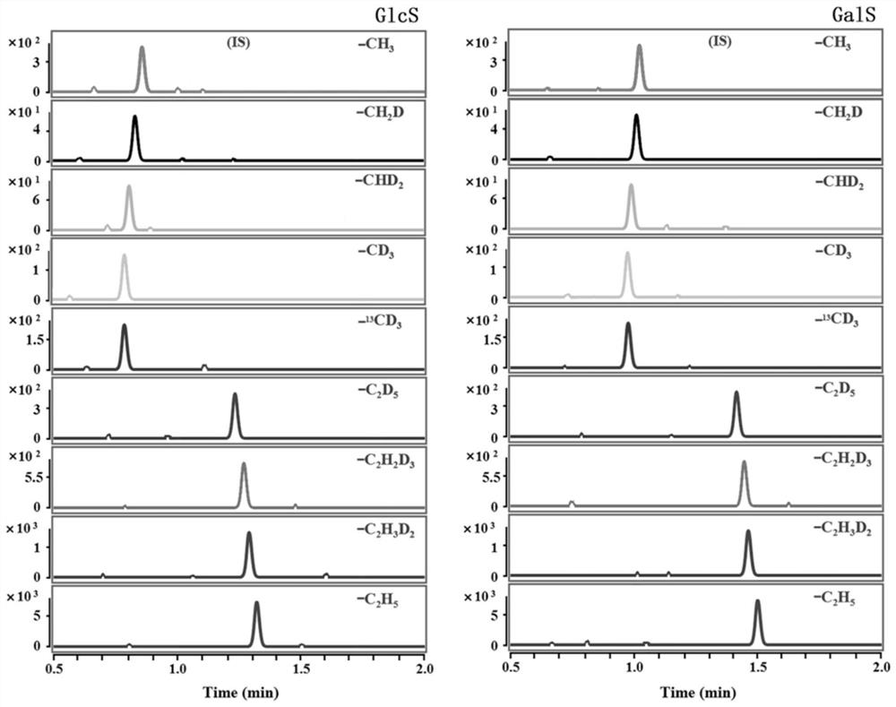
[0044] Chromatographic separation and qualitative and quantitative analysis of GlcS and GalS spiked samples in delipidated plasma:

[0045] GlcS and GalS were prepared in acetonitrile to obtain 1×10 -6 Mo / L standard stock solution, and then prepared with fat-free plasma to obtain concentrations of 0.02 / 0.02, 0.2 / 0.2, 2.0 / 2.0, 20.0 / 20.0, 50.0 / 50.0, 100.0 / 100.0, 400.0 / 400.0, 800.0 / 800.0 The concentration range of the mixed standard solution of GlcS and GalS in nmol / L covers the concentration range of GlcS / GalS in the plasma of patients with Gaucher disease or Krabbe disease. CH 3 / CH 2 D / CHD 2 / 13 cd 3 / CD 3 / C 2 h 5 / C 2 h 3 D. 2 / C 2 h 2 D. 3 / C 2 D. 5 -ALSF was dissolved in acetonitrile to give 2.5×10 -8mol / L derivatization reagent acetonitrile solution; take 50 μL mixed standard solution from 8 parts of delipidated plasma mixed standard solution and a single part of acetonitrile mixed standard solution in a 1.5 mL centrifuge tube, add 300 μL H 3 BO 3 -Na ...

Embodiment 2

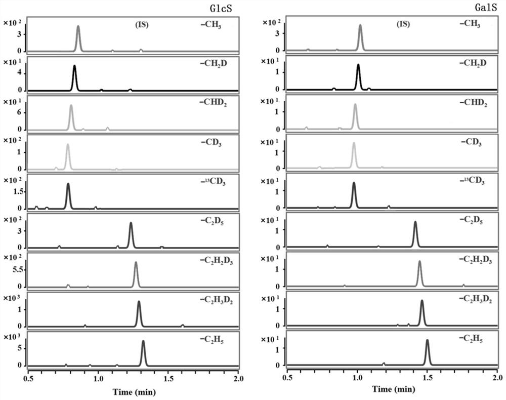
[0050] The detection and analysis of GlcS and GalS in the serum of a simulated Gaucher patient includes the following steps:

[0051] Take 8 healthy human serum samples from a local hospital, mix them evenly in a centrifuge tube, and centrifuge quickly at 4°C, take 500 μL of serum samples and add 500 μL methanol to a 1.5 mL centrifuge tube, vortex for 1.5 minutes and centrifuge to remove For protein precipitation, take 40 μL and place it in a 1.5 mL centrifuge tube, prepare 8 copies, and then add 10 μL of GlcS diluted with acetonitrile at a concentration of 25.0, 50.0, 100.0, 300.0, 500.0, 1000.0, 4000.0 nmol / L to each of the seven copies The concentration range of the standard solution after adding the standard covers the GlcS content range in the serum of patients with Gaucher disease. Add 10 μL of acetonitrile solution to the remaining portion as a control. In addition, take 50 μL of the mixed standard acetonitrile solution (from a concentration of 1×10 -8 mol / L GalS, the...

Embodiment 3

[0053] The detection of GlcS and GalS in the plasma of a simulated Gaucher patient includes the following steps:

[0054] Take 8 samples of healthy human plasma from a local hospital and mix them evenly in test tubes containing ethylenediaminetetraacetic acid (EDTA), and centrifuge them quickly at 4°C. Take 2 mL of plasma samples and add 8 mL of methanol into a 15.0 mL centrifuge tube, vortex Spin and shake for 2 minutes and centrifuge to remove protein precipitates, take 40 μL and put it in a 1.5mL centrifuge tube, prepare 8 copies, and then add 10 μL to each of the seven copies with concentrations of 25.0, 50.0, 100.0, 300.0, 500.0, 1000.0, 4000.0 GlcS standard solution diluted in nmol / L acetonitrile, the concentration range of the added solution covered the GlcS content range in the plasma of patients with Gaucher disease, and the remaining part was added with 10 μL acetonitrile solution as a control. In addition, take 50 μL of the mixed standard acetonitrile solution (from...


PUM


Abstract

The invention belongs to the technical field of analytical chemistry, in particular to a multi-channel mass spectrometry derivatization reagent for detecting sphingosine glucoside (GlcS) and sphingosine galactoside (GalS) and its preparation method and application, in particular to the design Synthesize and utilize CH 3 / CH 2 D / CHD 2 / 13 cd 3 / CD 3 / C 2 h 5 / C 2 h 3 D. 2 / C 2 h 2 D. 3 / C 2 D. 5 ‑Piperazinelevofloxacin‑N‑hydroxysuccinimidyl formate was used as a multi-channel mass spectrometry derivatization reagent for the simultaneous separation and quantitative analysis of GlcS and GalS in 8 biological samples. The structural formula of the derivative reagent is:. The invention designs and synthesizes a multi-channel mass spectrometry derivatization reagent for the first time. The derivatization reaction conditions are mild, the reaction speed is fast, the derivative product is stable, and the chromatographic separation of analytes and mass spectrometry detection sensitivity, accuracy and analysis throughput are significantly improved.

Description

technical field [0001] The invention belongs to the technical field of analytical chemistry, in particular to a multi-channel mass spectrometry derivatization reagent for simultaneous derivation, separation and detection of sphingosine glucoside (GlcS) and sphingosine galactoside (GalS) and its preparation method and application, Especially when it comes to designing and synthesizing and utilizing CH 3 / CH 2 D / CHD 2 / 13 cd 3 / CD 3 / C 2 h 5 / C 2 h 3 D. 2 / C 2 h 2 D. 3 / C 2 D. 5 -piperazine-levofloxacin-N-hydroxysuccinimide formate (CH 3 / CH 2 D / CHD 2 / 13 cd 3 / CD 3 / C 2 h 5 / C 2 h 3 D. 2 / C 2 h 2 D. 3 / C 2 D. 5 -ALSF) as a multi-channel mass spectrometry derivatization reagent for the simultaneous separation and quantitative analysis of GlcS and GalS in 8 biological samples. Background technique [0002] GlcS and GalS are glycosphingolipids in which the 1-hydroxyl group of sphingosine is bound to galactose and glucose, respectively. GlcS can ...

Claims


Application Information

Patent Timeline
Patent Type & Authority Patents(China)
IPC IPC(8): C07D498/06G01N30/02G01N30/06G01N30/32G01N30/34G01N30/72
CPCC07D498/06G01N30/02G01N30/06G01N30/32G01N30/34G01N30/72G01N2030/062G01N2030/324
Inventor 赵先恩陈世恩朱树芸
Owner 武汉科益研创科技有限公司
Learn More