Bladder cancer detection device and method
A technology for bladder cancer cells and cancer cells, which is applied in the field of bladder cancer detection devices and methods, and can solve the problems of increasing sample analysis time and cost, reducing background, etc.
- Summary
- Abstract
- Description
- Claims
- Application Information
AI Technical Summary
Problems solved by technology
Method used
Image
Examples
Embodiment 1-96
[0107] Example 1- Preparation of a 96-well plate plasma polymer substrate
[0108] For cell capture, 96-well plates (Corning, Costar) and microscope grade glass slides (Objekttrager) are used as solid substrates. Use silicon wafers for plasma polymer film thickness measurement.
[0109] Before plasma deposition, the glass slides and silicon wafers were washed in piranha solution (H2O2:H2SO4, 1:3), and rinsed thoroughly with milliQ ultrapure water. Use a sterile 96-well plate as obtained.
[0110] The oxazoline-based thin film coating is deposited on the solid substrate by continuous plasma deposition as described previously. (Macgregor-Ramiasa et al. 2015b; Ramiasa et al. 2015) Simply put, the customized parallel plate plasma reactor is placed in a vacuum (2.10 -2 mbar), and inoculate 2-methyl-2-oxazoline precursor (Sigma-Aldrich, Australia) with a needle valve in the chamber until a constant monomer flow rate of 8 standard cubic centimeters per minute (sccm) is reached. Then the ...
Embodiment 2
[0113] Example 2-Preparation of Microfluidic Plasma Polymer Substrate
[0114] The method described in Example 1 was used to form a PPOx-coated microscope slide. Then use standard techniques on the PPOx coated microscope slide and Ibidi IV 0.4 sticky (DSKH) Micro-channels are formed between the sticky-tear glass slides.
Embodiment 3
[0115] Example 3-Covalent attachment of antibody to plasma polymer substrate
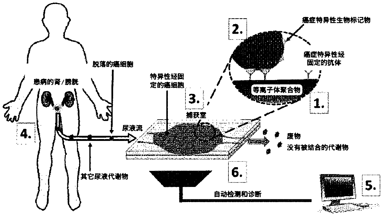
[0116] The cell capture "chamber" consists of individual wells (Example 1) or microchannels (Example 2) in a standard 96-well plate. The unique reactivity of plasma-deposited polyoxazoline and carboxylic acid is used to irreversibly bind antibodies to the plasma polymer substrate. (Macgregor-Ramiasa et al. 2015a; Schmidt et al. 1994; Tillet et al. 2011) Dissolve Anti-EpCAM antibody in 100% PBS at 10 μg.mL-1, gently pipette 50 μL onto the plasma polymer substrate and make They were combined overnight at 4°C. Then draw the antibody solution and pass in 0.1 mg.mL -1 The surface of the active polymer was blocked with skim milk by incubating for 15 minutes in the solution. Rinse the substrate completely with PBS. ToF SIMS analysis was used to confirm the immobilization of the antibody on the PPOx substrate.
PUM
 Abstract
Description
Claims
Application Information
 - R&D
- Intellectual Property
- Life Sciences
- Materials
- Tech Scout
- Unparalleled Data Quality
- Higher Quality Content
- 60% Fewer Hallucinations
Browse by: Latest US Patents, China's latest patents, Technical Efficacy Thesaurus, Application Domain, Technology Topic, Popular Technical Reports.
© 2025 PatSnap. All rights reserved.Legal|Privacy policy|Modern Slavery Act Transparency Statement|Sitemap|About US| Contact US: help@patsnap.com


