
Pulmonary function imaging processing method based on magnetic resonance imaging (MRI) technology

A technology of imaging processing and lung function, applied in medical science, sensors, diagnostic recording/measurement, etc., can solve the problem of improving the reproducibility and repeatability of pulmonary ventilation function maps and perfusion maps, the inability to combine objective indicators, pulmonary ventilation and Address issues such as fuzzy perfusion information to achieve the effect of promoting application

Pending Publication Date: 2020-01-07
CHINA JILIANG UNIV
View PDF4 Cites 1 Cited by
  • Summary
  • Abstract
  • Description
  • Claims
  • Application Information

AI Technical Summary

Problems solved by technology

[0006] However, there are several basic problems in the use of FD-MRI method for pulmonary function measurement: the measured lung ventilation and perfusion information is vague and cannot be clearly combined with the objective indicators of clinical pulmonary function tests; the measured pulmonary ventilation function map and perfusion map The reproducibility and reproducibility of the lung function need to be improved; it is necessary to design a fast acquisition method for magnetic resonance data suitable for pulmonary function testing, and to further improve the contrast and resolution of lung ventilation and perfusion maps

Method used


Image

  • Pulmonary function imaging processing method based on magnetic resonance imaging (MRI) technology
  • Pulmonary function imaging processing method based on magnetic resonance imaging (MRI) technology
  • Pulmonary function imaging processing method based on magnetic resonance imaging (MRI) technology

Examples


Embodiment Construction

[0035] Preferred embodiments of the present invention will be described in detail below with reference to the accompanying drawings, but the present invention is not limited to these embodiments. The present invention covers any alternatives, modifications, equivalent methods and schemes made within the spirit and scope of the present invention.

[0036] In order to provide the public with a thorough understanding of the present invention, specific details are set forth in the following preferred embodiments of the present invention, but those skilled in the art can fully understand the present invention without the description of these details.

[0037]In the following paragraphs the invention is described more specifically by way of example with reference to the accompanying drawings. It should be noted that all the drawings are in simplified form and use inaccurate scales, which are only used to facilitate and clearly assist the purpose of illustrating the embodiments of th...


PUM


Abstract

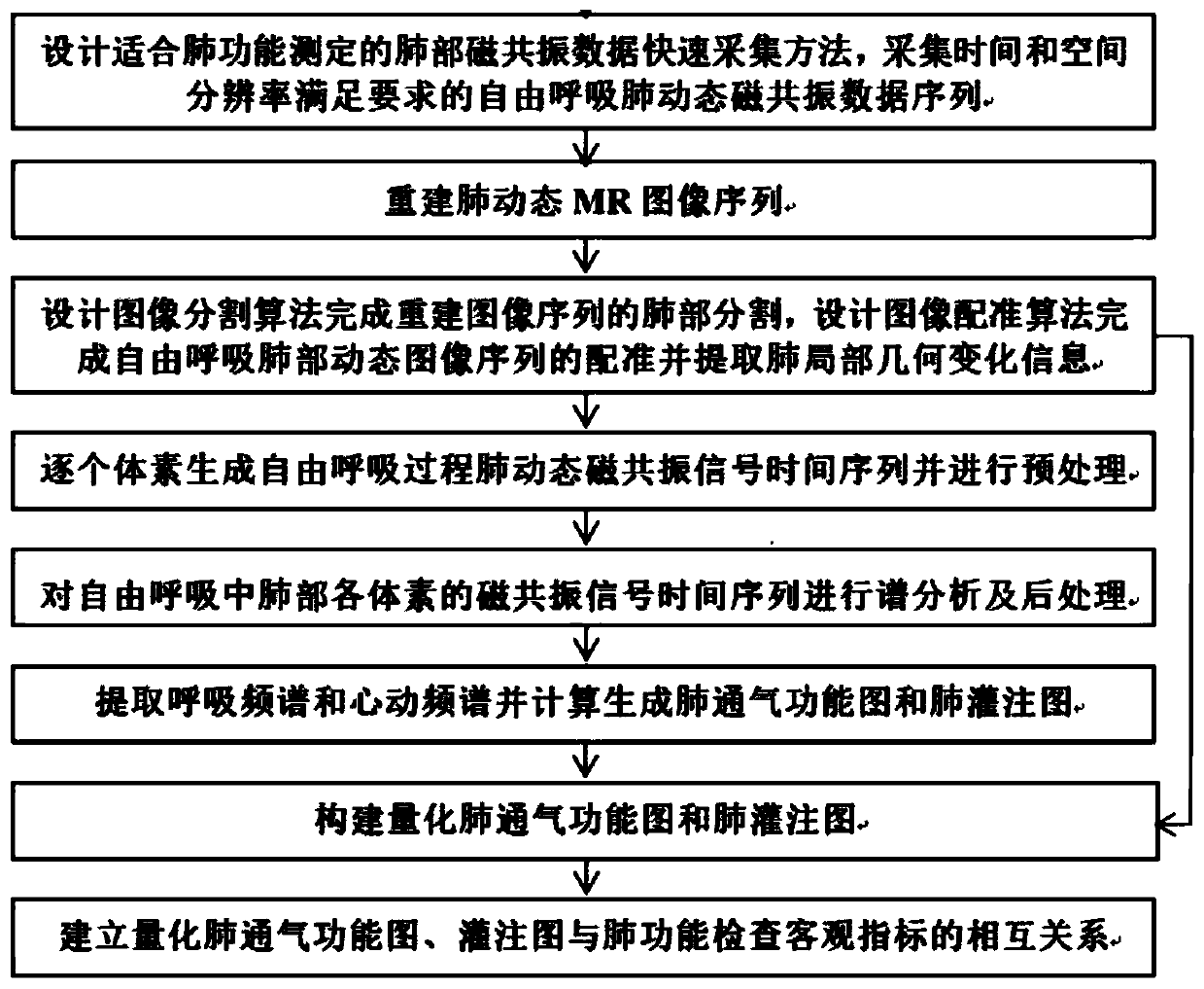
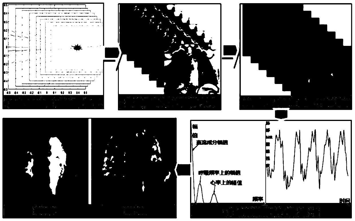
The invention discloses a pulmonary function imaging processing method based on magnetic resonance imaging (MRI) technology. The pulmonary function imaging processing method based on the MRI technology comprises the following steps: step one, rapidly acquiring dynamic magnetic resonance (MR) data series of free breathing lungs; step two, reconstructing dynamic MR image sequences of the lungs withgood spatial resolution and contrast by utilizing the MR data series acquired in the step one and carrying out MR image reconstruction; step three, carrying out segmentation of lung tissue of the MR image and registration of dynamic MR image sequences of the free breathing lungs so as to extract geometric information of lung tissue during respiratory movement; step four, building time series one by one according to the registered dynamic MR image sequences of the lungs, and carrying out spectral analysis; and step five, constructing quantitative pulmonary ventilation function map and pulmonaryperfusion map according to the registered geometric change information of the lung tissue during the respiratory movement in combination with structural information and quantitative MR image parameters of the lungs. The pulmonary function imaging processing method based on the MRI technology disclosed by the invention is safe, reliable, and capable of acquiring sensitive and accurate quantitativepulmonary ventilation function maps and pulmonary perfusion maps.

Description

technical field [0001] The present invention relates to the technical field, and more specifically, relates to a lung function imaging processing method based on MRI technology. Background technique [0002] Respiratory diseases such as chronic obstructive pulmonary disease and asthma are one of the main diseases that cause death. There are nearly 100 million chronic obstructive pulmonary disease (COPD) patients in China, and the incidence rate of people over 40 years old is as high as 13.7%. The main feature of this type of respiratory system disease is airway obstruction or tissue damage and airflow limitation. Lung volume, lung ventilation and ventilation function are the main objective analysis indicators. Pulmonary function instruments such as spirometer and plethysmograph are often used clinically. Determination. These measurement methods only reflect the overall function of the lung, but cannot reveal the local ventilation and ventilation function of the lung and its...

Claims


Application Information

Patent Timeline
IPC IPC(8): A61B5/055
CPCA61B5/055
Inventor 刘晓芳姜远才张纠郑峰
Owner CHINA JILIANG UNIV
Learn More