
Methods for visualization and 3D printing of multimodal cardiac images

A 3D printing, multi-modal technology, applied in the field of image processing

Active Publication Date: 2020-06-30
杭州海睿博研科技有限公司 +1
View PDF8 Cites 0 Cited by
  • Summary
  • Abstract
  • Description
  • Claims
  • Application Information

AI Technical Summary

Problems solved by technology

While these methods are adequate for the analysis of normal heart disease, there are considerable challenges in translating these images to understand the 3D spatial relationships occurring within the diseased heart as well as other organs of interest

Method used


Image

  • Methods for visualization and 3D printing of multimodal cardiac images
  • Methods for visualization and 3D printing of multimodal cardiac images
  • Methods for visualization and 3D printing of multimodal cardiac images

Examples


Embodiment Construction

[0049] In the following description, the present invention is described with reference to various examples. One skilled in the art will recognize, however, that the various embodiments may be practiced without one or more of the specific details, or with other alternative and / or additional methods, materials, or components. In other instances, well-known structures, materials, or operations are not shown or described in detail so as not to obscure aspects of the various embodiments of the invention. Similarly, for purposes of explanation, specific quantities, materials and configurations are set forth in order to provide a thorough understanding of embodiments of the invention. However, the invention may be practiced without these specific details. Furthermore, it should be understood that the various embodiments shown in the drawings are illustrative representations and are not necessarily drawn to scale.

[0050] In this specification, reference to "one embodiment" or "the...


PUM


Abstract

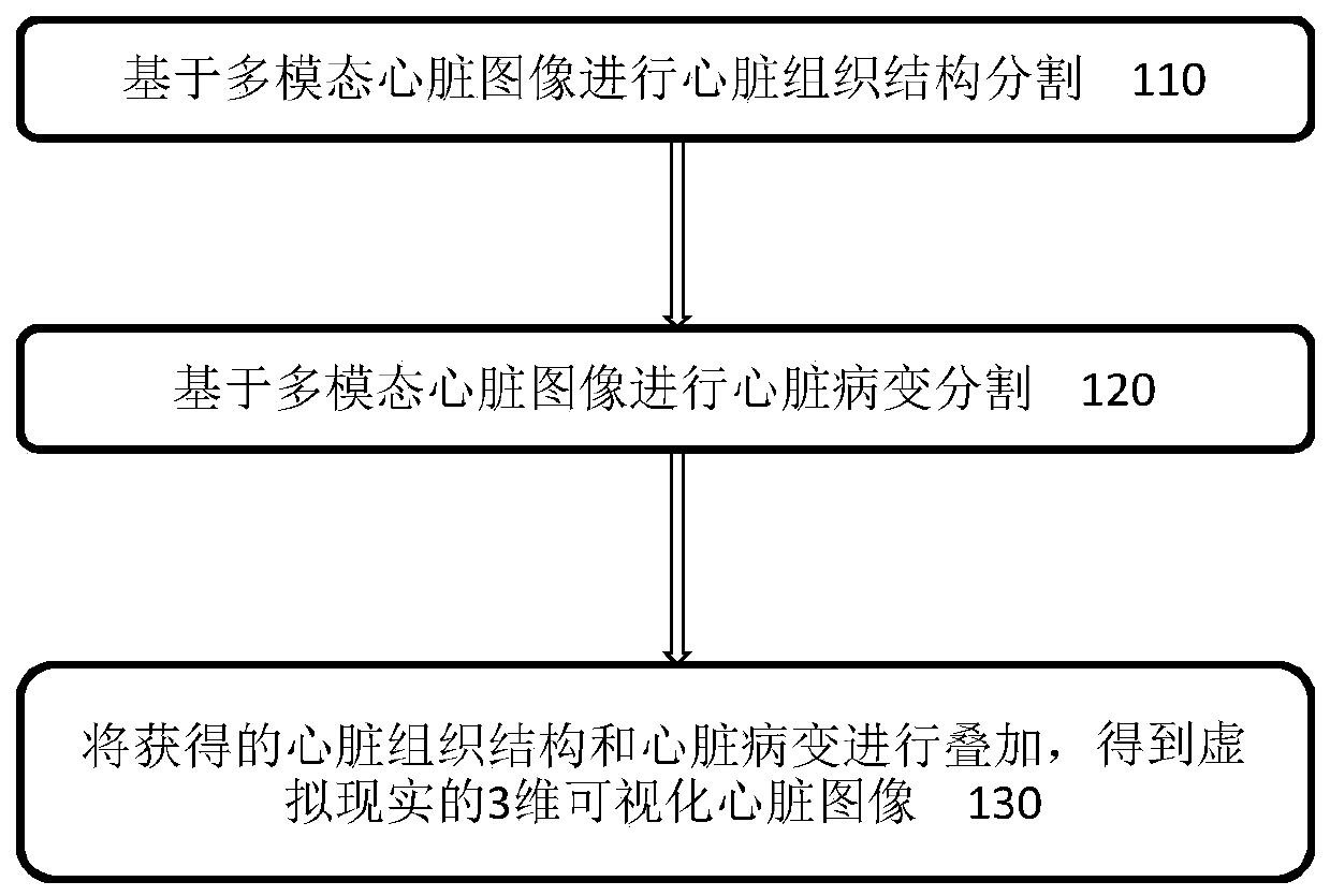
The invention discloses a multimodal cardiac image visualization method, comprising: segmenting cardiac tissue structure based on the multimodal cardiac image; segmenting cardiac lesion based on the multimodal cardiac image; and combining the obtained cardiac tissue structure and cardiac lesion Superimpose to obtain a three-dimensional visualization heart image in virtual reality; and control the 3D printer through the automatic analysis of the heart tissue structure and its lesions, use elastomeric materials to print the chambers and valves of the 3D heart and combine them with mixed reality for visual display.

Description

technical field [0001] The present invention relates to the field of image processing, and more specifically, the present invention relates to methods for visualization, 3D printing and mixed reality display of multimodal cardiac images. Background technique [0002] In 2015, there were 422.7 million cardiovascular disease patients worldwide, of whom 17.92 million died from cardiovascular disease. Cardiac magnetic resonance imaging has important clinical implications for assessing ventricular function, including calculation of left ventricular end-diastolic and end-systolic volumes. In calculating these volumes, it is usually necessary to demarcate the left ventricular myocardium. Therefore, accurate segmentation of the left ventricular endocardium is a prerequisite for evaluating left ventricular function, and manual segmentation is a long and tedious process. It takes 20 minutes for a doctor to manually segment a group of cases. [0003] In order to reduce the workload o...

Claims


Application Information

Patent Timeline
Patent Type & Authority Patents(China)
IPC IPC(8): G06T7/00G06T7/11G06T17/00G06K9/62G06N3/04G06N3/08B29C64/393B33Y50/02
CPCG06T7/0012G06T7/11G06T17/00G06N3/08B29C64/386B33Y50/00G06T2207/30048G06T2207/30096G06T2207/20081G06T2207/20084G06N3/045G06F18/2415G06F18/241
Inventor 杨光牛张明陆纬江荧辉李劳王承嘉叶旭炯董豪姜敏
Owner 杭州海睿博研科技有限公司
Learn More