Method and system for identifying grid change holes in wide-area fundus images based on deep learning

A deep learning, fundus image technology, applied in the field of medical image processing, can solve the problems of inability to detect and evaluate, time-consuming inspection, visual impairment, etc., and achieve the effect of reducing training expenditure, efficient disease evaluation, and reducing burden

Active Publication Date: 2022-03-11
SUN YAT SEN UNIV +1
View PDF0 Cites 0 Cited by
  • Summary
  • Abstract
  • Description
  • Claims
  • Application Information

AI Technical Summary

Problems solved by technology

Usually community hospitals or low-level general hospitals and physical examination centers do not have professional ophthalmologists, so they cannot conduct comprehensive detection and evaluation of such lesions
[0004] 2. The amount of information contained in the wide-area fundus image is about 4 times that of the traditional fundus image. The peripheral retina has a variety of grid-like degeneration and holes, and the early features are not obvious. Accurate interpretation of this fundus image requires ophthalmologists to undergo professional training and comparison. Accumulation of long-term experience
[0005] 3. Manual inspection of the peripheral retina requires mydriasis and the close cooperation between the fundus doctor and the patient. Most elderly patients or children do not cooperate well. The inspection takes a long time, and it is easy to miss the diagnosis of the disease due to insufficient cooperation.
[0006] In summary, due to the limitations of ophthalmologists' examination techniques in primary hospitals, and the time-consuming detailed inspection of peripheral retinal lattice degeneration and holes, it is difficult to carry out screening of peripheral retinal lattice degeneration and holes in large-scale population, which can lead to the failure of most patients. Delay in diagnosis and treatment, some patients have retinal detachment due to the progress of the disease, resulting in visual damage, and the visual damage caused by the disease is irreversible, which has caused great losses to individuals, families and society

Method used


Image

  • Method and system for identifying grid change holes in wide-area fundus images based on deep learning
  • Method and system for identifying grid change holes in wide-area fundus images based on deep learning
  • Method and system for identifying grid change holes in wide-area fundus images based on deep learning

Examples


Embodiment 1

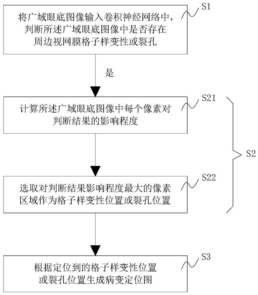
[0041] Such as figure 1 As shown, the present embodiment provides a method for identifying grid change holes in a wide-area fundus image based on deep learning, including the following steps:

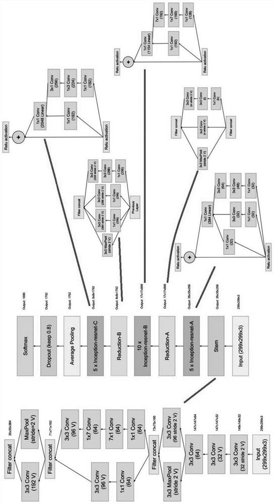
[0042] S1. Input the wide-area fundus image into the convolutional neural network, and judge whether there is lattice degeneration or hole in the peripheral retina in the wide-area fundus image;

[0043] S2. When it is judged that there is lattice degeneration or hole in the peripheral retina in the wide-area fundus image, the location of the lattice degeneration or the hole in the wide-area fundus image is located using a significant area algorithm.

[0044] In step S1, the convolutional neural network can be used to accurately and efficiently analyze the wide-area fundus image to determine whether there is a lesion on the image; in step S2, the salient area algorithm (Saliency Map) can be used to locate the lesion on the image. This can assist ophthalmologists to interpret patients' ...

Embodiment 2

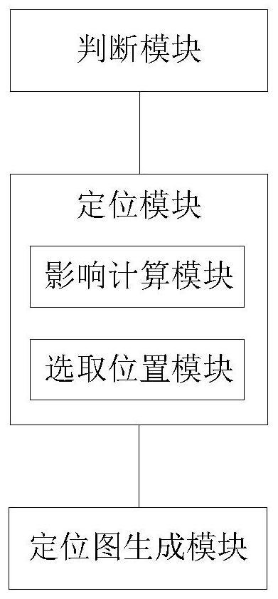
[0073] like image 3 As shown, this embodiment provides a system for identifying grid change holes in wide-area fundus images based on deep learning, including:

[0074] A judging module, configured to input the wide-area fundus image into the convolutional neural network, and judge whether there is a lattice-like degeneration or hole in the peripheral retina in the wide-area fundus image;

[0075] The positioning module is used to locate the grid-like degeneration or hole in the wide-area fundus image by using the salient area algorithm when it is judged that there is a grid-like degeneration or hole in the surrounding retina in the wide-area fundus image;

[0076] The judging module can accurately and efficiently analyze the wide-area fundus image through the convolutional neural network, and judge whether there is a lesion on the image; the positioning module can locate the location of the lesion on the image through the salient area algorithm (Saliency Map), which can assi...


PUM


Abstract

The present invention relates to a method and system for identifying lattice degeneration holes in a wide-area fundus image based on deep learning. The wide-area fundus image is input into a convolutional neural network to determine whether there is a lattice degeneration of the surrounding retina in the wide-area fundus image or not. Hole: when it is judged that there is lattice-like degeneration or hole in the peripheral retina in the wide-area fundus image, the location of the lattice-like degeneration or hole in the wide-area fundus image is located using the significant region algorithm. The invention can assist ophthalmologists to more accurately and conveniently interpret wide-area fundus images of patients.

Description

technical field [0001] The present invention relates to the technical field of medical image processing, and more specifically, to a method and system for identifying grid change holes in wide-area fundus images based on deep learning. Background technique [0002] The disadvantages of existing inspection techniques for peripheral retinal lattice degeneration and holes are high requirements for inspectors, time-consuming and labor-intensive inspections, and the possibility of implementing large-scale crowd screening for peripheral retinal lattice degeneration or holes is low. The main reasons are as follows: [0003] 1. Screening for peripheral retinal lattice degeneration or holes requires high technical requirements for ophthalmologists and requires years of clinical experience in fundus diseases. Usually, community hospitals or low-level general hospitals and physical examination centers do not have professional ophthalmologists, so they cannot conduct comprehensive dete...

Claims


Application Information

Patent Timeline
Patent Type & Authority Patents(China)
IPC IPC(8): A61B3/12A61B3/14
CPCA61B3/12A61B3/14
Inventor 林浩添李中文郭翀张凯林铎儒
Owner SUN YAT SEN UNIV
Why Eureka
Try Eureka