
Image analysis method, computer device and storage medium

An analysis method and image technology, applied in the medical field, can solve problems such as low efficiency, and achieve the effect of ensuring accuracy and improving efficiency

Inactive Publication Date: 2019-10-01
SHANGHAI UNITED IMAGING INTELLIGENT MEDICAL TECH CO LTD
View PDF6 Cites 6 Cited by
  • Summary
  • Abstract
  • Description
  • Claims
  • Application Information

AI Technical Summary

Problems solved by technology

[0005] Based on this, it is necessary to provide an image analysis method, computer equipment and storage medium for the technical problem of low efficiency in the method of diagnosing diseases based on chest radiographs in the above-mentioned prior art

Method used


Image

  • Image analysis method, computer device and storage medium
  • Image analysis method, computer device and storage medium
  • Image analysis method, computer device and storage medium

Examples


Embodiment Construction

[0051] In order to make the purpose, technical solution and advantages of the present application clearer, the present application will be further described in detail below in conjunction with the accompanying drawings and embodiments. It should be understood that the specific embodiments described here are only used to explain the present application, and are not intended to limit the present application.

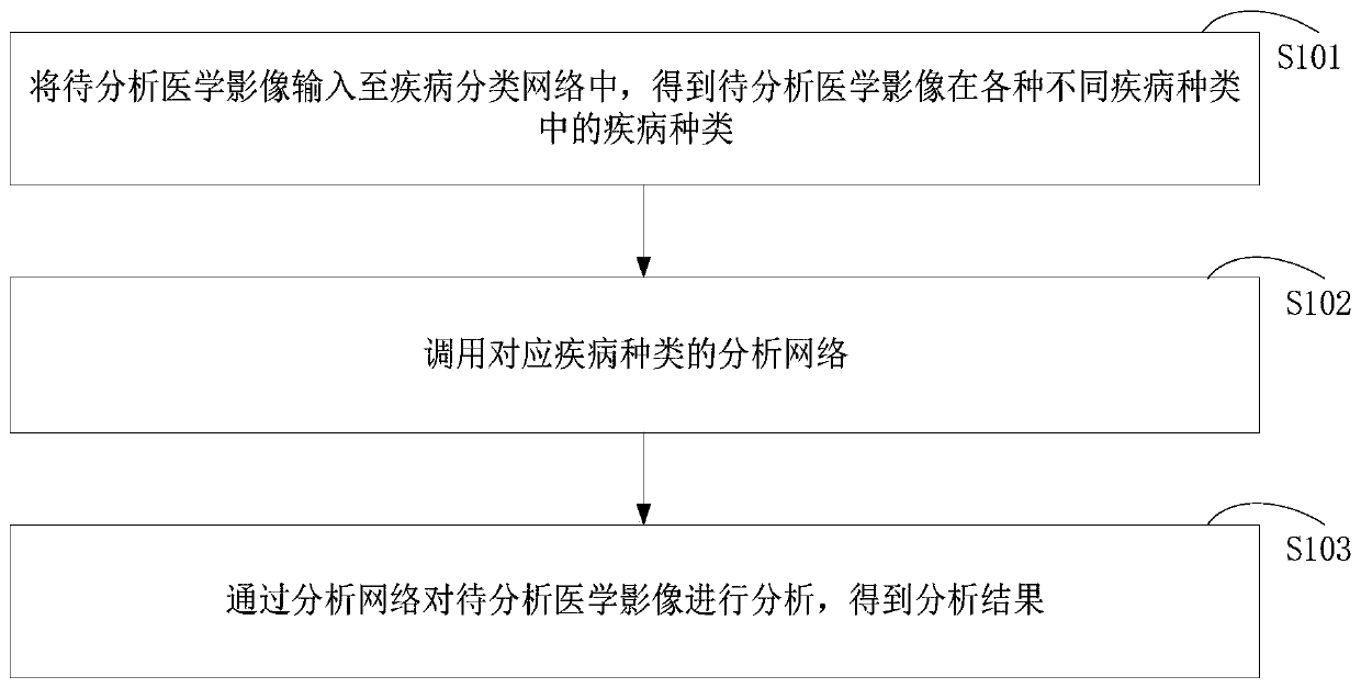
[0052] An image analysis method provided in the embodiment of this application can be applied to such as figure 1 In the application environment shown, the computer device includes a processor, a memory, a network interface and a database connected through a system bus. Wherein, the processor of the computer device is used to provide calculation and control capabilities. The memory of the computer device includes a non-volatile storage medium and an internal memory. The non-volatile storage medium stores an operating system, computer programs and databases. The internal...


PUM


Abstract

The invention relates to an image analysis method, a computer device and a storage medium. The whole process of analyzing the medical image from the analysis of the disease type of the medical image to be analyzed, the call to the corresponding disease type analysis network, and the analysis network to be analyzed is automatically performed by the computer device, and the whole process can be obtained without manual intervention. The analysis result of the medical image to be analyzed. Thus, the application of the method for diagnosing the disease according to the chest radiograph can not onlygreatly improve the efficiency of analyzing the medical image to be analyzed, but also ensure the accuracy of the analysis result.

Description

technical field [0001] The present application relates to the field of medical technology, in particular to an image analysis method, computer equipment and storage media. Background technique [0002] Because X-ray photography is convenient, simple, and economical, it becomes the preferred choice for chest examination. [0003] Chest film imaged by X-ray photography is an important means widely used in clinical diagnosis, for example, it can be used to check the thorax (including ribs, thoracic spine, soft tissue, etc.), thoracic cavity, lung tissue, mediastinum, heart and other diseases. Usually, when diagnosing chest diseases based on chest X-rays, doctors need to read the X-rays for each case and give a diagnosis report, or the doctor first reads the X-rays and then classifies the diseases, and then inputs the X-rays into the analysis software corresponding to the disease type , to get the corresponding diagnosis result. [0004] However, the method for diagnosing dise...

Claims


Application Information

Patent Timeline
IPC IPC(8): G06T7/00G06T7/11G06K9/62
CPCG06T7/0012G06T7/11G06T2207/10116G06T2207/20081G06T2207/20084G06T2207/30004G06F18/241
Inventor 何志强郑介志
Owner SHANGHAI UNITED IMAGING INTELLIGENT MEDICAL TECH CO LTD
Learn More