Detecting method for detecting methylation of specific gene

A technology of specific gene and detection method, applied in the field of detection of specific gene methylation, can solve the problems of high professional requirements of operators, cumbersome operation process, expensive fluorescent PCR equipment, etc., to achieve good sensitivity and specificity, and results. Ease of Analysis, Enhanced Sensitivity

Active Publication Date: 2019-09-27
天津康博尔生物基因技术有限公司
View PDF4 Cites 2 Cited by
  • Summary
  • Abstract
  • Description
  • Claims
  • Application Information

AI Technical Summary

Problems solved by technology

However, this type of method requires relatively expensive fluorescent PCR equipment, and the operation process is relatively cumbersome. The test cycle of the workflow execution takes about 8 hours, and the professional requirements of the operators are relatively high, so it cannot well meet the actual screening needs.

Method used


Image

  • Detecting method for detecting methylation of specific gene
  • Detecting method for detecting methylation of specific gene
  • Detecting method for detecting methylation of specific gene

Examples


Embodiment 1

[0029] Example 1: Preparation of lateral flow nucleic acid detection test paper

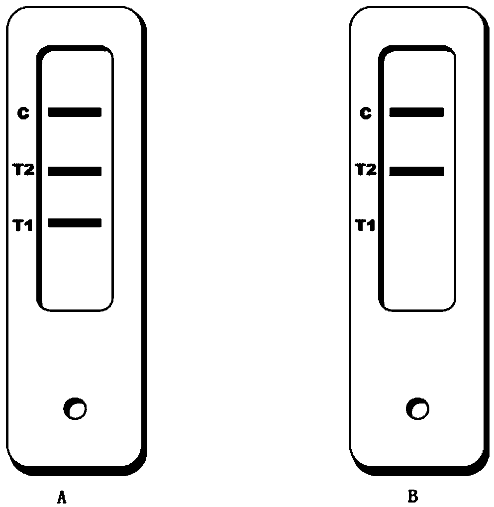
[0030] see attached Figure 1A , wherein, SP: sample pad CP: binding pad NM: nitrocellulose membrane detection area AP: absorbent pad.

[0031] (1) Preparation of nitrocellulose membrane detection area:

[0032] The nitrocellulose detection membrane was cut into long strips, placed on the platform of the spotting apparatus, and digoxigenumab, FITC monoclonal antibody and biotin-BSA were sprayed on the detection membrane to form T1, T2 and C lines. Dry at 42°C for 30min or air dry at room temperature.

[0033] (2) Preparation of the binding pad:

[0034] Cut the glass wool into long strips, place them on the platform of the spray point meter, take the nanoparticle markers, add a certain volume of pretreatment solution, spray them on the glass wool, and dry at 50°C for 30 minutes.

[0035] (3) Preparation of sample pad:

[0036] The glass wool was cut into long strips, soaked in a certain volum...

Embodiment 2

[0039] Example 2: Treatment of DNA with Sulfite Solution

[0040] (1) Extract human genomic DNA using a commercial DNA extraction kit or other suitable methods;

[0041] (2) Take 10 μL of DNA solution (100ng-2 μg) and 90 μL of sulfite solution, mix well, centrifuge briefly, and place them in a PCR instrument to react;

[0042] (3) reaction conditions are as follows:

[0043] The first stage: 95 ℃ 5min, 1 cycle,;

[0044] The second stage: 95℃ for 30S, 70℃ for 10min, 15 cycles;

[0045] (4) Purify DNA using a commercial DNA purification kit or other suitable methods.

Embodiment 3

[0046]Example 3: Methylation-dependent restriction endonuclease treatment of DNA

[0047] (1) Extract human genomic DNA using a commercial DNA extraction kit or other suitable methods;

[0048] (2) Take 10μL of DNA solution (100ng-2μg), 5μL of 10x reaction buffer, 2U of methylation-dependent restriction endonuclease and 35μL of deionized water, mix well, centrifuge briefly, and place in a PCR machine to react;

[0049] (3) reaction conditions are as follows:

[0050] The first stage: 30 ℃ 1hr, 1 cycle,;

[0051] The second stage: 15min at 70°C, 1 cycle.


PUM


Abstract

The invention discloses a detecting method for detecting methylation of a specific gene. The detecting method comprises a methylation-specific PCR amplification reaction and side flow nucleic acid detection. The methylation of the specific gene is identified and amplified based on a PCR method; the methylation is detected by combining a side flow technology after an amplification product is marked. The detecting method for detecting the methylation of the specific gene has the beneficial effects that higher detecting sensitivity and specificity than those of similar technologies or products are realized, and the methylation of the specific gene is detected quickly, conveniently, sensitively and economically by adopting a simple and high-efficiency DNA methylation treatment technology and a PCR amplification technology, and utilizing nano-color latex microspheres as a visible signal of nucleic acid detection instead of optical signals, such as a fluorescent probe, without complicated operation and expensive instruments; the detecting method for detecting the methylation of the specific gene is suitable for popularization, application and industrialization.

Description

technical field [0001] The invention relates to the field of biological detection, in particular to a detection method for detecting methylation of a specific gene. Background technique [0002] Malignant tumor (cancer) is one of the diseases that seriously threaten human health. The death of malignant tumor accounts for about a quarter of all deaths of Chinese residents, and it is on the rise. One of the reasons is that 60%-80% of cancers in China are first diagnosed in the middle and late stages, and the five-year survival rate of patients is relatively low. Therefore, screening for cancer at an early stage is of great significance for cancer diagnosis and treatment. At present, the conventional screening methods for cancer are mainly tumor marker detection and imaging examination, but these methods are either not sensitive enough to lead to missed detection, or insufficient specificity to lead to misdiagnosis, or cause discomfort to patients or low cost performance, resu...

Claims


Application Information

Patent Timeline
Patent Type & Authority Applications(China)
IPC IPC(8): C12Q1/6858
CPCC12Q1/6858C12Q2531/113C12Q2523/125C12Q2565/625C12Q2521/301
Inventor 张亮杨光
Owner 天津康博尔生物基因技术有限公司
Why Eureka
Try Eureka