
Medical image scanning automatic positioning method based on deep learning

A deep learning and automatic positioning technology, applied in the field of automatic positioning of medical image scanning, can solve the problem of inability to achieve accurate coordinate positioning of multiple organs and parts, and achieve the effects of fast calculation, reduced radiation, and reduced repetition

Active Publication Date: 2019-09-10
浙江明峰智能医疗科技有限公司
View PDF14 Cites 26 Cited by
  • Summary
  • Abstract
  • Description
  • Claims
  • Application Information

AI Technical Summary

Problems solved by technology

This method is mainly aimed at delineating and classifying the target area of ​​tumor radiotherapy and organs at risk, and cannot realize the precise coordinate positioning of multiple organs and multiple parts.

Method used


Image

  • Medical image scanning automatic positioning method based on deep learning
  • Medical image scanning automatic positioning method based on deep learning
  • Medical image scanning automatic positioning method based on deep learning

Examples


Embodiment Construction

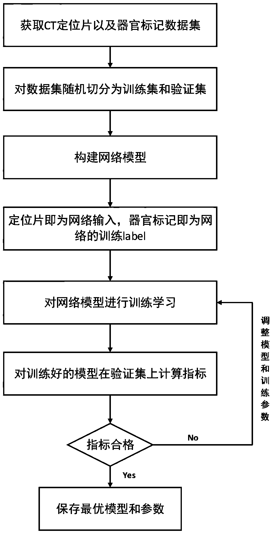
[0030] see figure 1 As shown, the medical image scanning automatic positioning method based on deep learning of the present invention comprises the following steps:

[0031] S1. Obtain a large number of positioning slice images, and randomly segment each category in the positioning slice images into a training set T, a verification set V and a test set U, and merge the images of the same three sets as a training set;

[0032] S2. According to the requirements of the deep learning target detection model, manually mark each organ that needs to be marked in each positioning slice image in the training set. The mark information includes the center coordinate information of the prediction frame, the length and width information of the prediction frame And the category information of the prediction box;

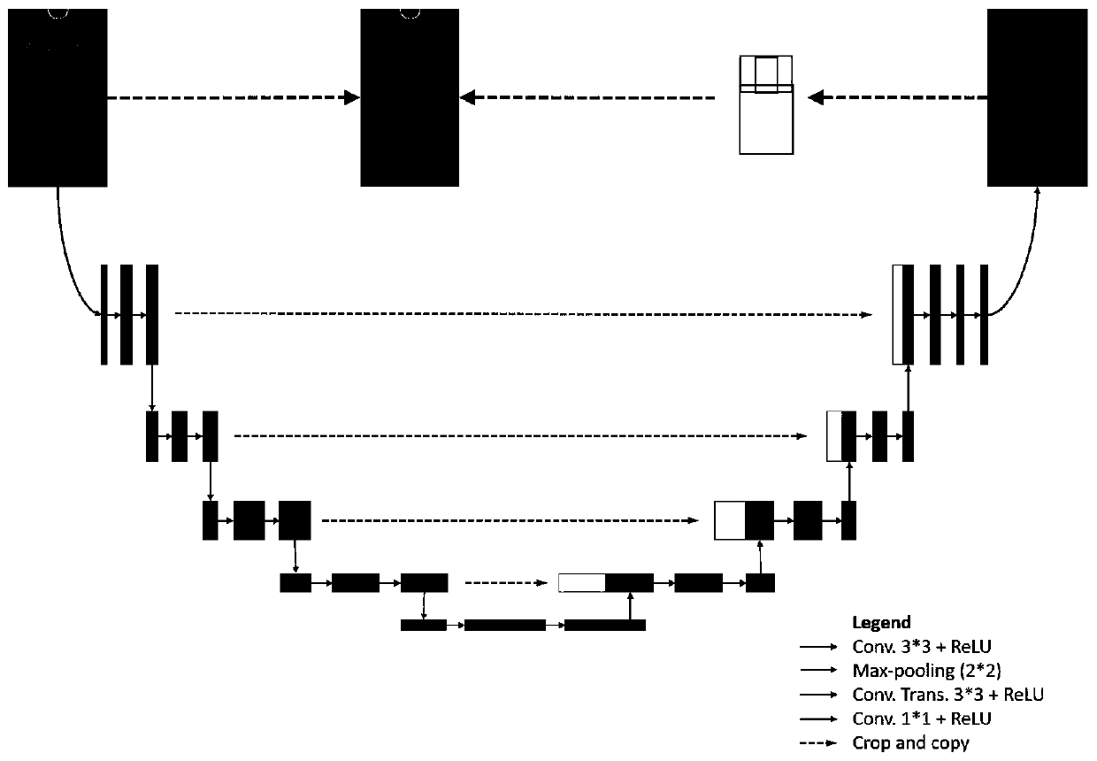
[0033] S3, construct the network model of deep learning, use the training set T and the verification set V as the input of the network model respectively, use the organ mark as th...


PUM


Abstract

The invention provides a medical image scanning automatic positioning method based on deep learning, and the method comprises the steps: obtaining a large number of positioning sheet images, and carrying out the random segmentation of the images into a training set, a verification set, and a test set; labeling each organ needing to be labeled in each positioning sheet image; constructing a deep learning network model, taking the training set and the verification set as input of the network model for training, and obtaining training parameters; operating the network model on the test set by using the training parameters; obtaining coordinates, width and height data and category data of a left upper point of a positioning frame of each organ needing to be examined, carrying out further post-processing on the obtained positioning frame data according to the requirements of CT and PET scanning parameters to obtain final positioning frame data of a human body part needing to be examined, and completing deep learning; and scanning the patient by using the network model which finishes deep learning, and finally obtaining an automatically positioned scanning image.

Description

technical field [0001] The invention relates to an automatic positioning method for medical image scanning. Background technique [0002] Computed Tomography (CT) scanning can obtain information on continuous layers of the human body. The helical scanning speed is fast, and it is not easy to miss lesion information. It has great flexibility in image processing and can obtain image layers in any direction. It has been widely used. It is used in three-dimensional imaging of human body, angiographic imaging, cardiac imaging, cerebral perfusion imaging and other fields. [0003] CT imaging uses X-rays to scan the parts of the human body for inspection. The detector receives the X-rays after passing through the human body and converts them into visible light, which is converted into electrical signals by photoelectric converters, and then converted into digital signals by analog-to-digital converters. After the computer image reconstruction program is reconstructed into a CT ima...

Claims


Application Information

Patent Timeline
Patent Type & Authority Applications(China)
IPC IPC(8): G06T7/73G06T7/62G06N3/04
CPCG06T7/73G06T7/62G06T2207/10081G06T2207/10104G06T2207/30004G06N3/045
Inventor 叶宏伟何宏炜徐怿弘
Owner 浙江明峰智能医疗科技有限公司
Learn More