
Blood vessel enhancement method of color fundus image

A fundus image and color technology, applied in image enhancement, image data processing, instruments, etc., can solve the problems of shock death, poor safety, limited application range, etc., and achieve the effect of good safety, high reliability, and wide application range

Active Publication Date: 2019-06-07
CENT SOUTH UNIV
View PDF3 Cites 5 Cited by
  • Summary
  • Abstract
  • Description
  • Claims
  • Application Information

AI Technical Summary

Problems solved by technology

However, with the use of fluorescein, some patients may have mild nausea and vomiting reactions, and individual patients may have allergic reactions and even shock death
Therefore, the existing fundus fluorescein angiography has very poor safety and extremely limited scope of application.

Method used


Image

  • Blood vessel enhancement method of color fundus image
  • Blood vessel enhancement method of color fundus image
  • Blood vessel enhancement method of color fundus image

Examples


Embodiment Construction

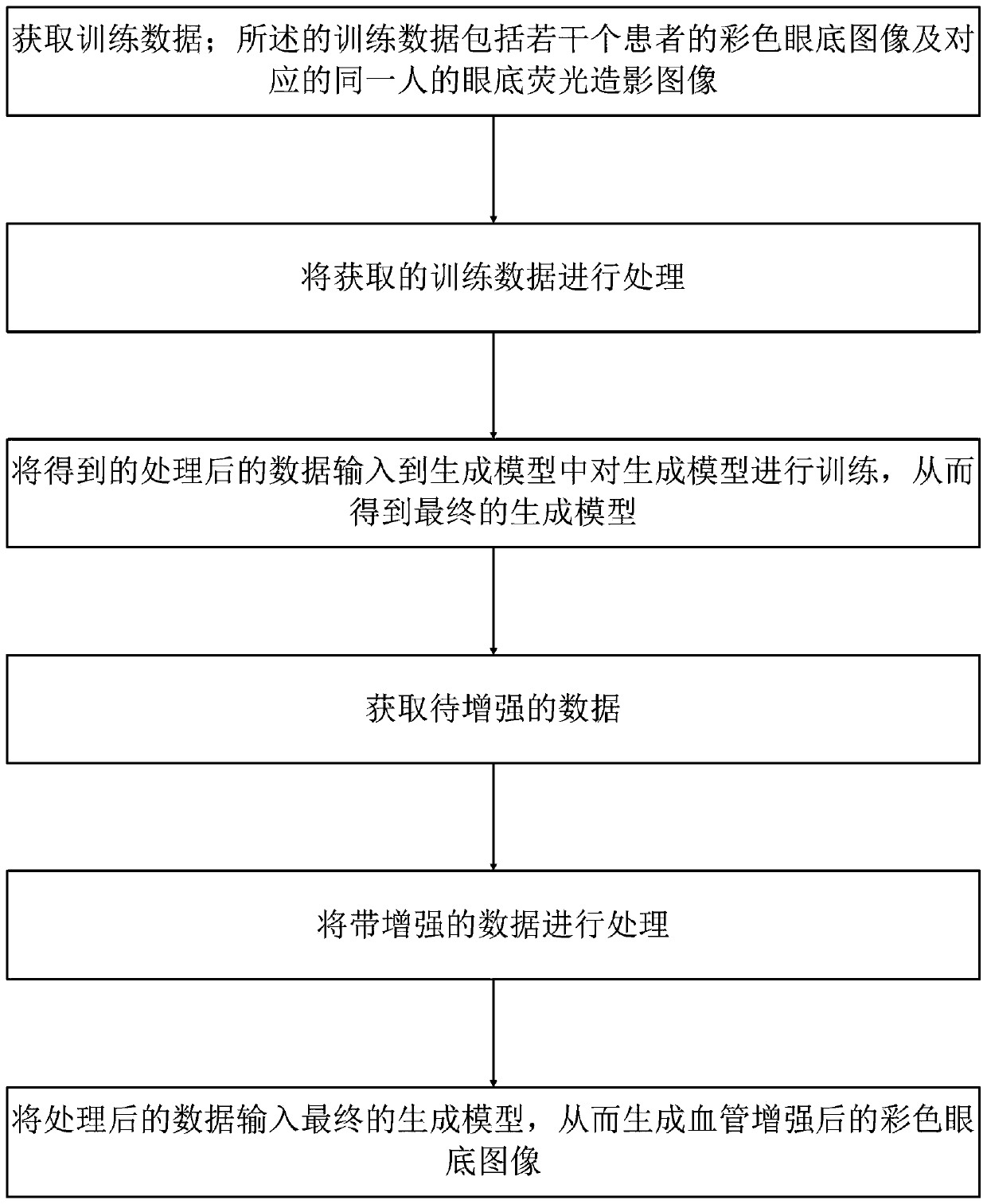
[0044] like figure 1 Shown is the method flowchart of the method of the present invention: the blood vessel enhancement method of the color fundus image provided by the present invention includes the following steps:

[0045] S1. Acquire training data; the training data includes color fundus images of several patients and corresponding fundus fluorescein contrast images of the same person;

[0046] S2. Process the training data obtained in step S1; specifically, the following steps are used for processing:

[0047] A. Normalize the training data obtained in step S1; use the following formula for normalization:

[0048]

[0049] where p(x,y) is the normalized pixel value of point (x,y), is the original pixel value of the point (x, y), P is the set of pixel values ​​of all points in the image, max(P) is the maximum value of the pixel value, and min(P) is the minimum value of the pixel value;

[0050] B. Cut the normalized training data; specifically, cut the normalized im...


PUM


Abstract

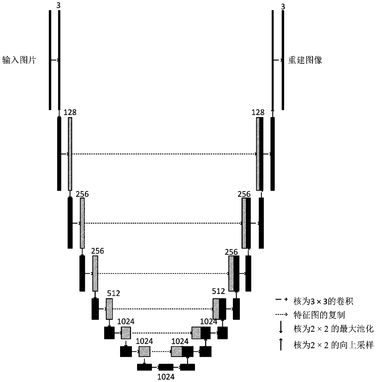
The invention discloses a blood vessel enhancement method for a color fundus image. The method comprises the steps of obtaining and processing training data; inputting the data into a generation modelto train the generation model and obtain a final generation model; acquiring and processing data to be enhanced; and inputting the data into a final generation model to generate a blood vessel enhanced color fundus image. According to the invention, the generation model is established; a deep neural network is used for learning blood vessel imaging characteristics of a fluorescence contrast image, information deeper than gray level textures and the like can be learned, the blood vessel enhancement effect of an eye fundus image is more remarkable, and a generated image can be effectively closer to a target image through design of a loss function; therefore, the method can effectively generate the blood vessel enhanced color fundus image according to the existing color fundus image, and ishigh in reliability, good in safety and wide in application range.

Description

technical field [0001] The invention specifically relates to a blood vessel enhancement method of a color fundus image. Background technique [0002] With the development of economy and technology and the improvement of people's living standards, people's requirements for health are getting higher and higher. With the emergence, development and popularization of intelligent algorithms, more and more intelligent algorithms are used in the field of medical assistance, which has brought great help to people's medical treatment. The analysis of retinal images, especially the accurate segmentation of retinal vessels and the analysis of retinal vascular structures, can assist in the screening of diabetic retinopathy. Therefore, the analysis of retinal images has been one of the research hotspots in recent years. [0003] The fundus vascular structure is usually acquired through retinal color fundus images taken by a fundus camera. However, due to the uneven illumination in the ...

Claims


Application Information

Patent Timeline
IPC IPC(8): G06T5/00
Inventor 邹北骥陈瑶朱承璋陈昌龙张子谦
Owner CENT SOUTH UNIV
Learn More