
A mammary gland ultrasonic image recognition and analysis method and system

An ultrasound image and analysis method technology, applied in image analysis, image data processing, neural learning methods, etc., can solve the problems of cumbersome lesion measurement process, difficulty in distinguishing the nature of lesions, and low recognition rate of breast lesions, and improve the accuracy of diagnosis. , the effect of reducing repeated operations and increasing the robustness of the model

Pending Publication Date: 2019-05-07
CHISON MEDICAL TECH CO LTD
View PDF4 Cites 32 Cited by
  • Summary
  • Abstract
  • Description
  • Claims
  • Application Information

AI Technical Summary

Problems solved by technology

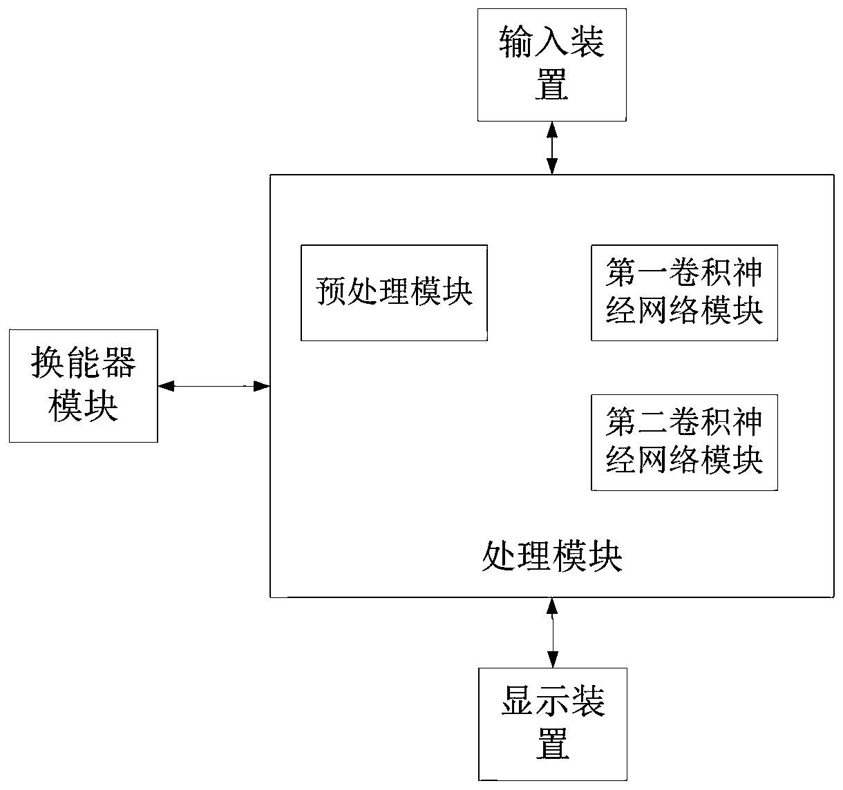
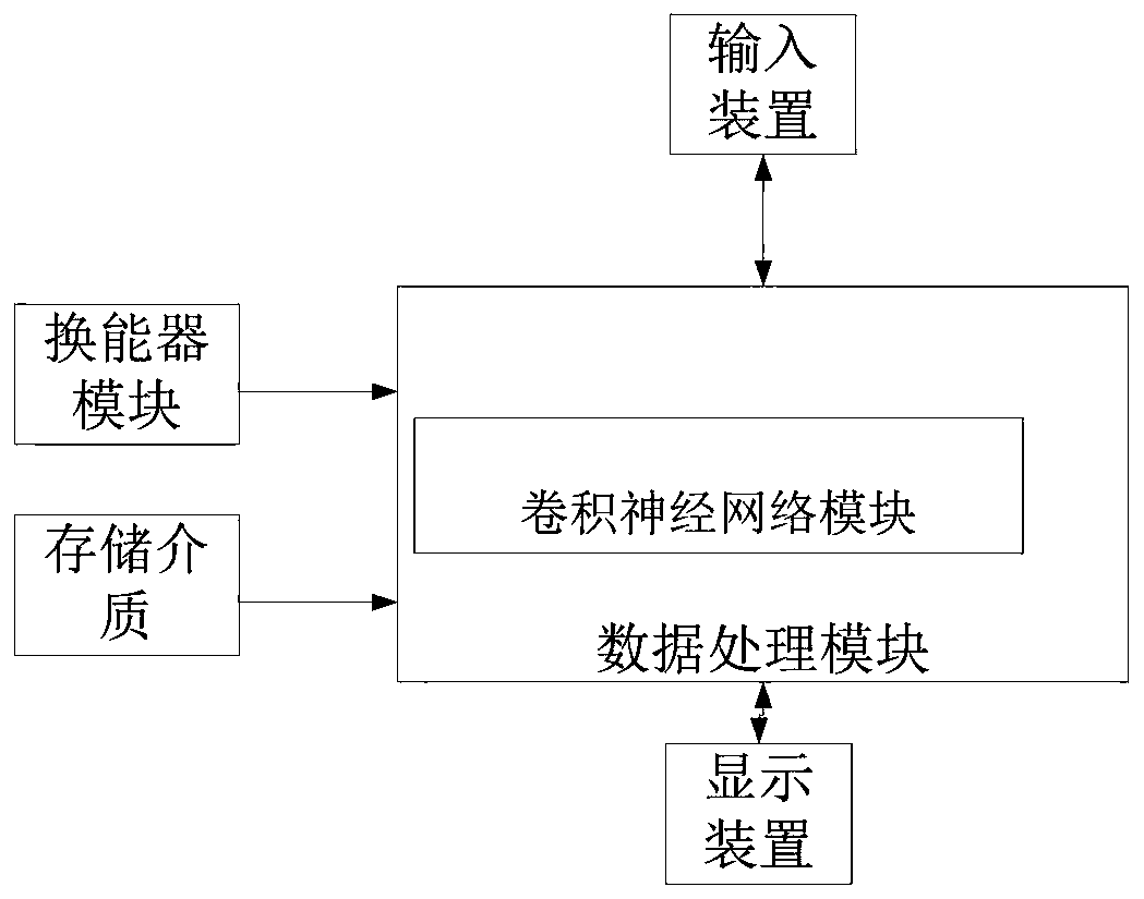
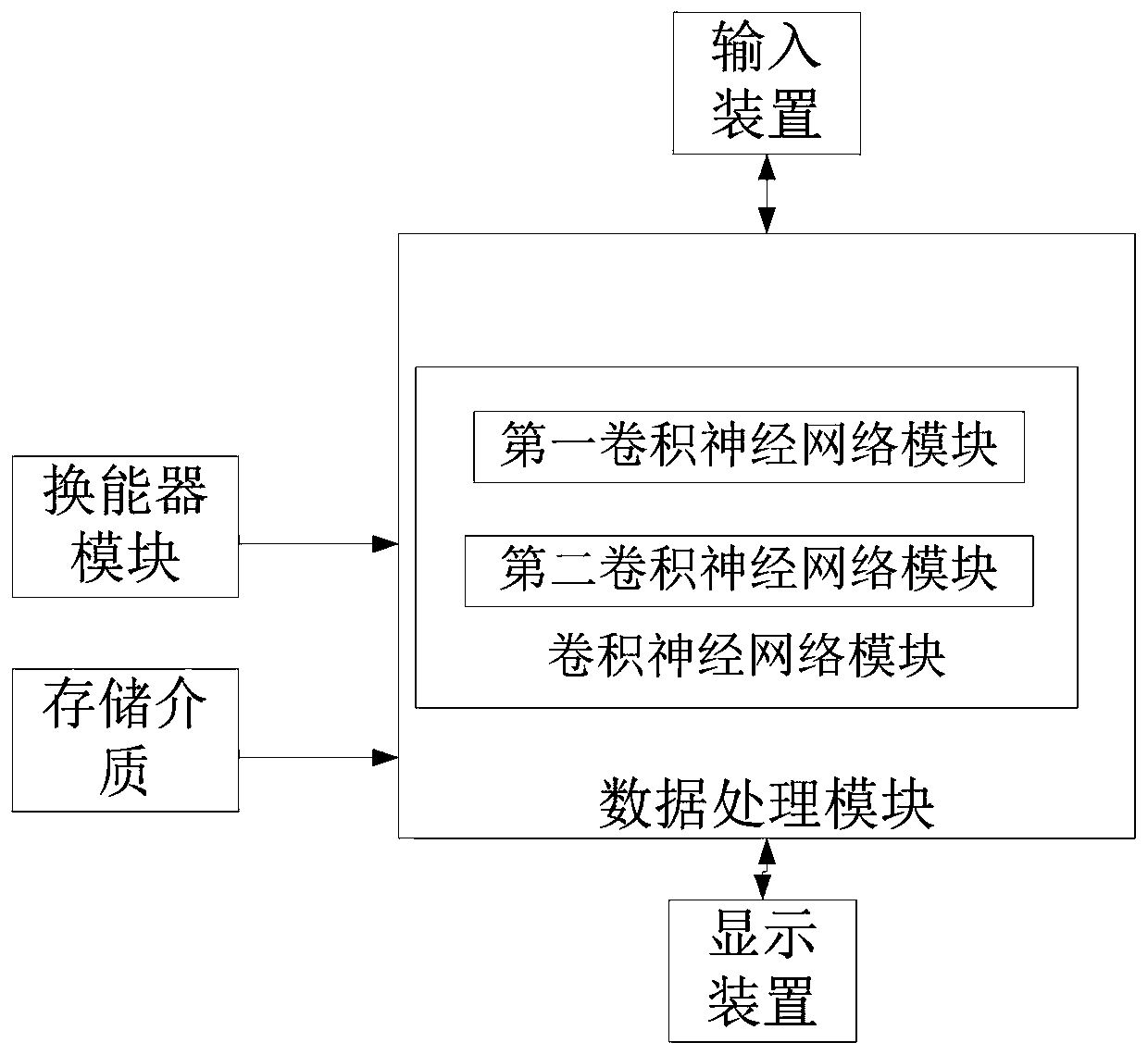
[0005] In order to solve the problems of low recognition rate of breast lesions, difficulty in distinguishing the nature of lesions, and cumbersome measurement process of lesions in the process of breast ultrasound examination and diagnosis, the present invention provides an automatic analysis of breast examination images, identification of breast lesions, property analysis and focus area contours Method for extraction, automatic measurement of parameters

Method used


Image

  • A mammary gland ultrasonic image recognition and analysis method and system
  • A mammary gland ultrasonic image recognition and analysis method and system
  • A mammary gland ultrasonic image recognition and analysis method and system

Examples


Embodiment Construction

[0043] The present invention will be further described below in conjunction with drawings and embodiments.

[0044] In the following examples, the terms "first" and "second" are used purely as labels, and are not considered to have numerical requirements for their modifiers.

[0045] A neural network "module" or "unit" as used herein means, but is not limited to, a software or hardware component that performs a specific task, such as a Field Programmable Gate Array (FPGA) or an Application Specific Integrated Circuit (ASIC) or a processor, such as a CPU , GPU. A module may advantageously be configured to reside on the addressable storage medium and configured to execute on one or more processors. Thus, by way of example, a module may include a component (such as a software component, an object-oriented software component, a class component, and a task component), a process, a function, an attribute, a procedure, a subroutine, a program code segment, a driver, firmware, microc...


PUM


Abstract

The invention provides a method and a system for automatically analyzing a mammary gland ultrasonic image, identifying a mammary gland focus and then carrying out focus area contour extraction and parameter calculation. According to the invention, the convolutional neural network is used to construct the recognition classification recognition and segmentation extraction model, the breast lesion area in the video or image can be recognized, the lesion area contour can be automatically extracted, and the parameters of the lesion area can be calculated. According to the method and the system, theauxiliary diagnosis accuracy is remarkably improved, repeated operations of doctors are greatly reduced, and the robustness is good.

Description

technical field [0001] The invention relates to the technical field of ultrasonic examination, in particular to a breast ultrasonic image recognition and analysis method and system. Background technique [0002] At present, when using ultrasound equipment to examine the patient's breast, the image quality of the machine is affected by the machine parameter settings, the angle of the probe during inspection, and the doctor's experience, which makes it difficult to quickly identify the lesions that appear on the image during the real-time examination of the patient. ; and due to the heavy workload of sonographers every day, affected by physical and emotional factors, it is difficult to accurately touch and analyze every lesion on all patients; Operations such as delineation and measurement of lesions in each lesion area found in , require a large number of button operations. At present, it is difficult for the equipment to conveniently and quickly obtain quantitative indicato...

Claims


Application Information

Patent Timeline
IPC IPC(8): G06T7/00G06T7/11G06T7/12G06T7/62G06N3/04G06N3/08
Inventor 甘从贵赵明昌莫若理
Owner CHISON MEDICAL TECH CO LTD
Learn More