Image segmentation method, device and equipment and storage medium
An image segmentation and image technology, applied in the field of medical image processing, can solve the problems of incomplete segmentation and segmentation leakage, and achieve the effect of ensuring integrity and suppressing data leakage.
- Summary
- Abstract
- Description
- Claims
- Application Information
AI Technical Summary
Problems solved by technology
Method used
Image

Examples
Embodiment 1
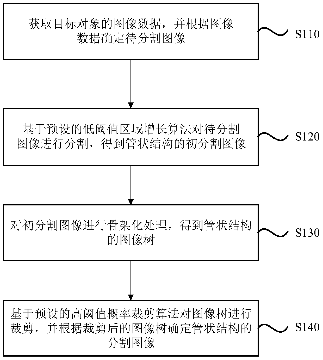
[0043] figure 1 It is a flowchart of an image segmentation method provided in Embodiment 1 of the present invention. This embodiment is applicable to the case of image segmentation with tubular structural features, especially suitable for the case of coronary artery image segmentation. The method can be executed by the image segmentation device provided in the embodiment of the present invention, and the device can be realized by software and / or hardware. see figure 1 , the method of the embodiment of the present invention specifically includes the following steps:
[0044] S110. Acquire image data of a target object, and determine an image to be segmented according to the image data.
[0045] Wherein, the image data of the target object can be obtained based on electronic computer X-ray tomography technology, which can be based on X-ray beams to scan a layer of a certain thickness of the target object, and the detector receives the X-rays that pass through this layer and c...
Embodiment 2
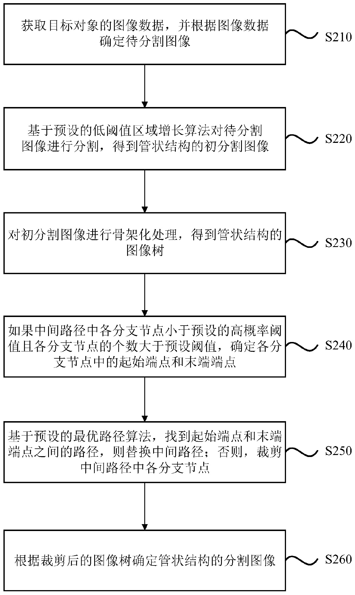
[0070] figure 2 It is a flowchart of an image segmentation method provided in Embodiment 2 of the present invention. This embodiment is optimized on the basis of the above technical solutions. In this embodiment, "cutting the image tree based on the preset high-threshold probability clipping algorithm" is specifically optimized as "if each branch node in the middle path is less than the preset high-probability threshold and the number of each branch node is greater than the preset Set the threshold to determine the start endpoint and end endpoint in each branch node, where the branch node is a node other than the root node and the leaf node; based on the preset optimal path algorithm, find the starting endpoint and the end endpoint path, replace the middle path; otherwise, cut each branch node in the middle path". Wherein, explanations of terms that are the same as or corresponding to the above embodiments are not repeated here. Correspondingly, such as figure 2 As shown...
Embodiment 3
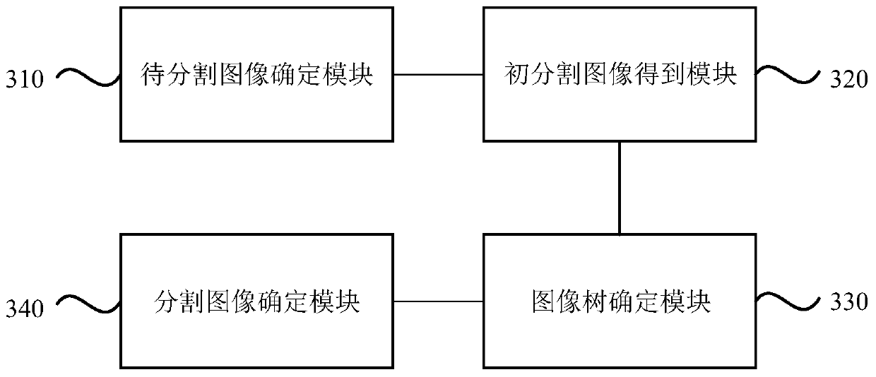
[0086] image 3 It is a structural block diagram of an image segmentation device provided in Embodiment 3 of the present invention, and the device is used to implement the image segmentation method provided in any of the above embodiments. The device and the image segmentation method of the above-mentioned embodiments belong to the same inventive concept. For the details not described in detail in the embodiment of the image segmentation device, reference may be made to the above-mentioned embodiment of the image segmentation method. see image 3 , the device may specifically include: an image to be segmented determination module 310 , an initial segmented image obtaining module 320 , an image tree determination module 330 and a segmented image determination module 340 .
[0087] Wherein, the image to be segmented determination module 310 is used to obtain image data of the target object, and determine the image to be segmented according to the image data;
[0088] The initi...
PUM
 Abstract
Description
Claims
Application Information
 - R&D
- Intellectual Property
- Life Sciences
- Materials
- Tech Scout
- Unparalleled Data Quality
- Higher Quality Content
- 60% Fewer Hallucinations
Browse by: Latest US Patents, China's latest patents, Technical Efficacy Thesaurus, Application Domain, Technology Topic, Popular Technical Reports.
© 2025 PatSnap. All rights reserved.Legal|Privacy policy|Modern Slavery Act Transparency Statement|Sitemap|About US| Contact US: help@patsnap.com