Dual-modal endoscope device based on liquid lens self-focusing

A liquid lens, self-focusing technology, applied to endoscopes, gastroscopes, esophagoscopes, etc., can solve the problems of limited imaging depth, low resolution and indistinguishable, lack of morphological structure information, etc. The effect of image distortion

Pending Publication Date: 2019-02-26
XIAMEN UNIV
View PDF0 Cites 4 Cited by
  • Summary
  • Abstract
  • Description
  • Claims
  • Application Information

AI Technical Summary

Problems solved by technology

However, in the existing multimodal endoscopy technologies such as fluorescence and photoacoustic endoscopy, although the fluorescence mode can realize the detection and imaging of fluorescence signals of early tissue lesions, it lacks morphological structure information and has low resolution to distinguish lesion groups. The fine features of the part; although the photoacoustic endoscopic microscope can improve the image resolution, but the imaging depth has a certain limit; at the same time, due to the surface inhomogeneity of the intestinal tissue, the focal point of the probe beam is defocused and the image distortion

Method used


Image

  • Dual-modal endoscope device based on liquid lens self-focusing
  • Dual-modal endoscope device based on liquid lens self-focusing
  • Dual-modal endoscope device based on liquid lens self-focusing

Examples


Embodiment Construction

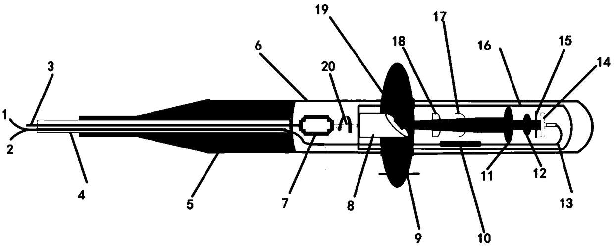
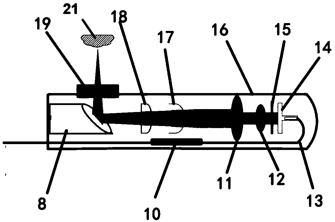
[0020] like figure 1 , figure 2 , image 3 As shown, this embodiment includes: single-mode fiber core 1, multi-mode fiber core 2, cable 3, optical fiber and cable housing 4, outer cavity body protective cover 5, endoscope outer cavity body 6, micro-motor 7, spherical surface Mirror 8, external cavity scanning window 9, fiber coupler 10, achromatic lenses 11 and 12, double-clad optical fiber 13, beam collimator 14, variable pinhole diaphragm 15, rotating scanning cavity 16, auto Focusing liquid lens 17, aspheric lens 18, main external cavity scanning window 19, rotating shaft 20, wherein: spherical mirror 8, achromatic lens 11 and 12, beam collimator 14, self-focusing liquid lens 17, double pack Layer fiber coupling 10 , double-clad fiber bundle 13 and aspheric lens 18 are located inside rotating scanning cavity 16 .

[0021] According to the principle of electrowetting, the self-focusing liquid lens 17 changes the curvature of the liquid mold and changes the focal length b...


PUM

PropertyMeasurementUnit
Focal lengthaaaaaaaaaa
Clear apertureaaaaaaaaaa
Thicknessaaaaaaaaaa

Abstract

The invention provides a dual-modal endoscope device based on liquid lens self-focusing. The dual-modal endoscope device based on liquid lens self-focusing comprises a shell and a rotary scanning cavity arranged in the shell and provided with a reflector. The rotary scanning cavity receives exciting light and outputs an OCE signal and a fluorescence signal reflected by a sample respectively through a single-mode optical fiber and a multi-mode optical fiber to achieve panoramic scanning. The OCE and FLE technologies are integrated, the structure and functions of the interior of an organism aresubjected to real-time high-resolution varifocal imaging, a precise and effective morphological structure information image is provided for early detection for precisely monitoring gastrointestinal tract system diseases, and meanwhile quantitative optical coherence and fluorescence two-dimensional and three-dimensional dual-modal images with high sensitivity and high specificity are given.

Description

technical field [0001] The present invention relates to a technology in the field of medical equipment, specifically a dual-mode endoscope device based on liquid lens self-focusing, which adopts double-clad optical fiber to realize high-sensitivity and high-specificity fluorescence endoscopic imaging, and at the same time It can realize automatic focusing optical coherent imaging of complex irregular structure organisms. It is an endoscope with automatic focusing characteristics that integrates two imaging modalities. Background technique [0002] At present, optical endoscopy has been commonly used in clinical disease detection. For example, fluorescence endoscopy uses exogenous fluorescent molecular probes to target target organisms to achieve molecular functional high-sensitivity imaging. Structural imaging, especially three-dimensional imaging of tissue tomography, has limitations. Optical coherence tomography (OCE), based on the measurement of optical weak coherent bac...

Claims


Application Information

Patent Timeline
IPC IPC(8): A61B5/00A61B1/00A61B1/04A61B1/273A61B1/31
CPCA61B1/00167A61B1/04A61B1/043A61B1/273A61B1/31A61B5/0066A61B5/0071A61B5/0084
Inventor 赵庆亮黄豆豆林文珍刘基嫣
Owner XIAMEN UNIV
Why Eureka
Try Eureka