
A kind of preparation method of biotype multilayer nerve conduit containing mesenchymal stem cells

A nerve conduit and mesenchymal stem cell technology, applied in the design and preparation of nerve conduits, can solve problems such as uncontrollable and unavailable nerve conduits, and achieve the effects of short time consumption, enhanced safety, and mild operating conditions

Active Publication Date: 2020-06-30
ZHEJIANG UNIV
View PDF2 Cites 0 Cited by
  • Summary
  • Abstract
  • Description
  • Claims
  • Application Information

AI Technical Summary

Problems solved by technology

However, the attachment position and distribution density of growth factors and neural stem cells on the nerve conduit are random and uncontrollable, and it is impossible to obtain neural conduits with uniform distribution, gradient distribution or programmed distribution of neural stem cells and nerve growth factors.

Method used


Image

  • A kind of preparation method of biotype multilayer nerve conduit containing mesenchymal stem cells
  • A kind of preparation method of biotype multilayer nerve conduit containing mesenchymal stem cells
  • A kind of preparation method of biotype multilayer nerve conduit containing mesenchymal stem cells

Examples


Embodiment 1

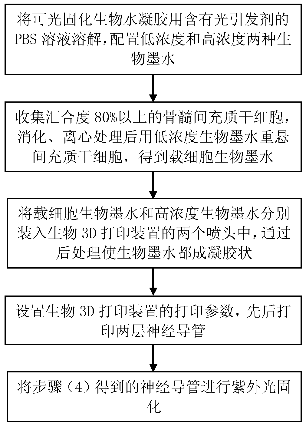
[0037] Such as figure 1 As shown, a method for preparing a biological multilayer nerve conduit containing mesenchymal stem cells, the specific steps are as follows:

[0038] (1) Weigh a certain mass of sterile GelMA into a 10ml centrifuge tube, dissolve it in PBS solution with 0.1% (w / v) LAP, and prepare 5% (w / v) and 30% (w / v) GelMA biological ink.

[0039] (2) Collect bone marrow mesenchymal stem cells (BMSCs) with cell confluence of 80% or more in the culture dish, digest the cells with 0.25% (w / v) trypsin solution, and then perform centrifugation, after digestion and centrifugation , centrifugation speed 100rpm, centrifugation time 5min. Pour off the supernatant in the centrifuge tube, use the 5% (w / v) GelMA bio-ink prepared in step (1) to resuspend the cells, gently pipette to make the cells evenly distributed in the bio-ink, and the cell density in the bio-ink is 1.0 *10 6 , to obtain 5% (w / v) GelMA bioink loaded with cells.

[0040] (3) Introduce the 5% (w / v) GelMA ...

Embodiment 2

[0048] Same as Example 1, the difference is that in step (1), the concentration of the configured low-concentration GelMA bio-ink is 15%, and other conditions remain unchanged. Prepare 6 double-deck nerve guides and test, the average value of the compression modulus of the inner layer of the nerve guide tube is 250KPa, the average value of the tensile modulus is 37.5KPa; the average value of the outer layer mechanical compression modulus is 700KPa, the tensile modulus The average value is 150KPa.

Embodiment 3

[0050] Same as Example 1, the difference is that in step (4), the two nozzles uniformly select 22G needles (inner diameter 0.413mm), and other conditions remain unchanged, resulting in a length of 50mm, an inner diameter of 3mm, an outer diameter of 4.652mm, and an inner diameter of 4.652mm. An initial double-layer hollow nerve conduit with a layer thickness of 0.413 mm and an outer layer thickness of 0.413 mm.

[0051] The final double-layer nerve guide was obtained after ultraviolet curing, and the mechanical compression and tensile modulus of the inner and outer layers of the obtained double-layer nerve guide were tested. Compared with Example 1, the test results remained basically unchanged.


PUM

PropertyMeasurementUnit
compressive modulusaaaaaaaaaa
thicknessaaaaaaaaaa

Abstract

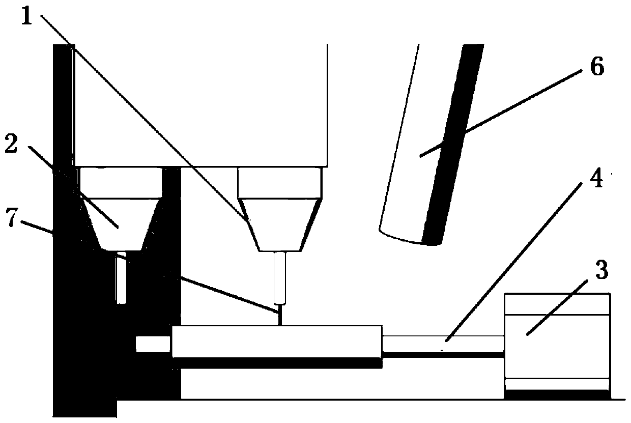
The invention discloses a preparation method of a biological multilayer nerve conduit containing mesenchymal stem cells. The method comprises the following steps: (1) dissolving a photocurable biological hydrogel with a PBS solution containing a photoinitiator to obtain two concentrations of biological ink; (2) collecting mesenchymal stem cells with confluence degree of more than 80%, and resuspending the mesenchymal stem cells with low concentration of biological ink after treatment to obtain cell-loaded biological ink; 3) load that cell-loaded biological ink and the high-concentration biological ink into two nozzles of the biological 3D printing device respectively; (4) setting printing parameters, firstly printing the cell-carrying biological ink on the optical axis rotating uniformly to obtain a first layer of nerve conduit; And then printing high-concentration biological ink to obtain a double-layer hollow tubular nerve conduit; (5) Biological multilayer nerve conduit was obtainedby ultraviolet irradiation. The invention has the advantages of short preparation time, simplicity, mild operation conditions, simple structure, good biocompatibility and degradability of the prepared nerve conduit.

Description

technical field [0001] The invention belongs to the field of design and preparation of nerve guides, in particular to a method for preparing biological multilayered nerve guides containing mesenchymal stem cells. Background technique [0002] Nervous system (nervous system) is the system that plays a leading role in the regulation of physiological functions and activities in the body. It is mainly composed of nervous tissue and is divided into two parts: the central nervous system and the peripheral nervous system. Various traumas such as compression, stretching, tearing, etc. and other factors such as tumors and local ischemia can easily cause damage to the nervous system, resulting in loss of nervous system function and other neurological diseases, seriously reducing the quality of work and life of patients. The current mainstream nerve repair techniques are as follows: direct anastomosis, autologous or allogeneic nerve transplantation, and nerve conduit repair. Direct an...

Claims


Application Information

Patent Timeline
Patent Type & Authority Patents(China)
IPC IPC(8): C12N5/077A61L27/40A61L27/22A61L27/38A61L27/52A61L27/58A61L27/50
CPCA61L27/222A61L27/3834A61L27/50A61L27/52A61L27/58A61L2430/32
Inventor 尹俊俞梦飞刘婧懿严梦玲傅建中吴俊杰
Owner ZHEJIANG UNIV
Learn More