An MRI brain tumor image segmentation method based on dbn neural network

An image segmentation and neural network technology, applied in the field of MRI brain tumor image segmentation based on DBN neural network, can solve problems such as unrealistic and poor scalability, and achieve improved detection rate, enhanced robustness, and enhanced segmentation accuracy. Effect

Active Publication Date: 2021-03-09
XIDIAN UNIV
View PDF6 Cites 0 Cited by
  • Summary
  • Abstract
  • Description
  • Claims
  • Application Information

AI Technical Summary

Problems solved by technology

However, this type of method often needs to manually extract features in advance, so it requires the designer to have relevant professional knowledge, which is not realistic in many cases, and the manually extracted features have the disadvantages of strong pertinence and poor scalability.

Method used


Image

  • An MRI brain tumor image segmentation method based on dbn neural network
  • An MRI brain tumor image segmentation method based on dbn neural network
  • An MRI brain tumor image segmentation method based on dbn neural network

Examples


Embodiment Construction

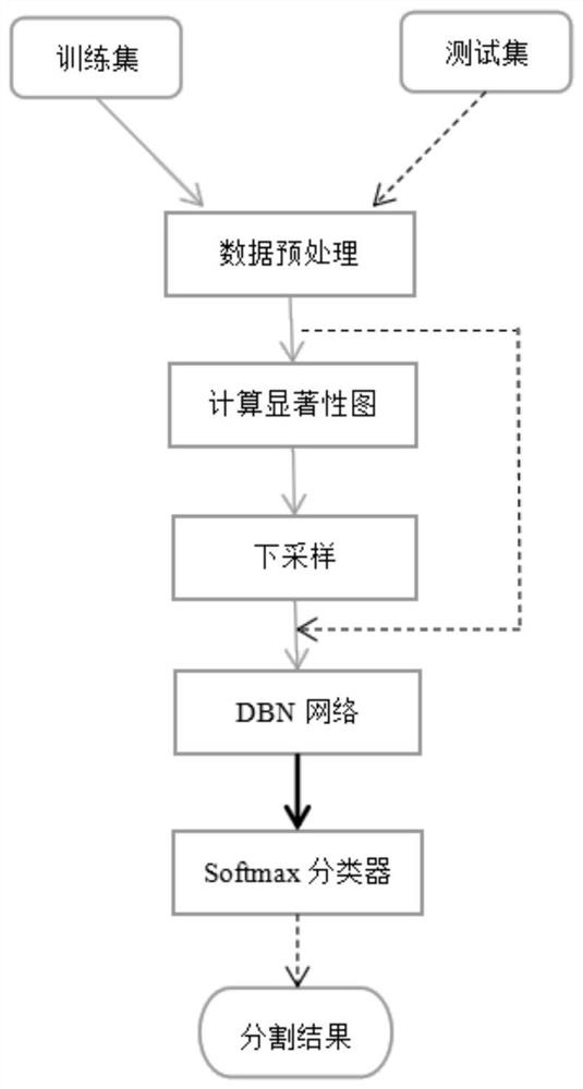
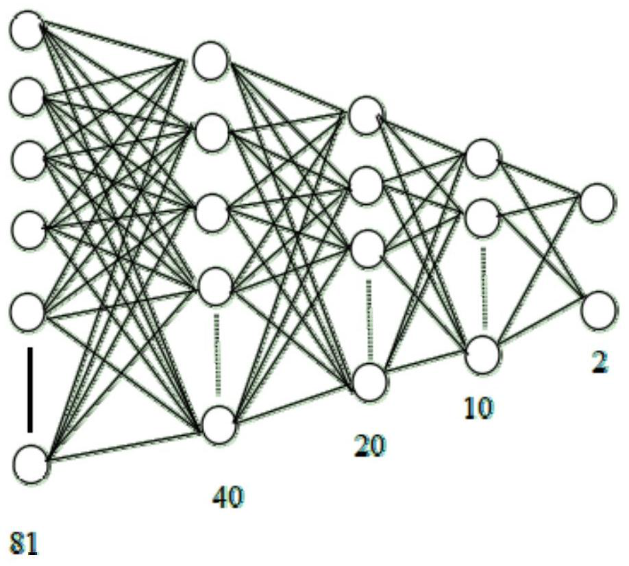
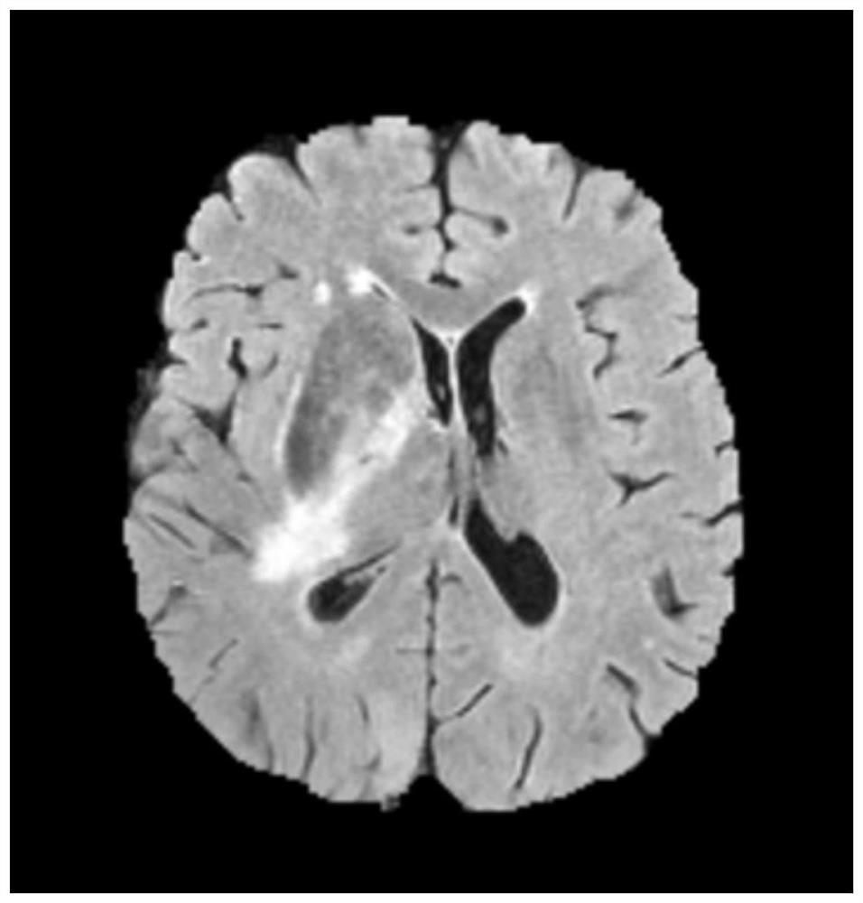
[0057] The invention provides a method for segmenting MRI brain tumor images based on a DBN neural network, which can be used to assist doctors in diagnosing and segmenting brain tumors. The realization process is as follows: firstly select multiple images from the existing patient brain MRI sequence image database as training samples, preprocess them and calculate the saliency map. Then the downsampling is sent to the DBN neural network for unsupervised and supervised training successively. After the training is completed, the test image to be segmented can be sent to the network for segmentation, and finally the segmentation result is output. The present invention extracts image features by means of deep learning, eliminating the tediousness and instability of manually extracting features. In addition, the introduction of downsampling to balance samples and a visual attention mechanism improves the accuracy of pixel classification, leading to better segmentation results for...


PUM


Abstract

The invention discloses an MRI brain tumor image segmentation method based on a DBN neural network. First, a plurality of images are selected from an existing patient brain MRI sequence image database as training samples, preprocessed and a saliency map is calculated; and then The downsampling is sent to the DBN neural network for unsupervised and supervised training successively. In view of the extremely unbalanced training samples, the non-tumor area is downsampled to improve the detection rate of positive samples; after the training is completed, you can The test image to be segmented is sent to the network for segmentation, and the visual attention model is introduced to enhance the network's segmentation accuracy for difficult-to-segment areas, and finally output the segmentation results.

Description

technical field [0001] The invention belongs to the technical field of image processing, and in particular relates to a method for segmenting MRI brain tumor images based on a DBN neural network. Background technique [0002] In recent years, brain tumors have become one of the tumors with the highest morbidity and mortality. Magnetic resonance imaging (MRI) can perform high spatial resolution and high contrast imaging of brain soft tissue, and is the best choice for doctors to analyze brain structure, so it is widely used clinically. In the brain MRI image processing, the precise segmentation of the tumor part is a crucial step, which plays a vital role in the doctor's subsequent analysis and judgment. At present, this step still relies heavily on manual segmentation, which is very time-consuming and has strong instability. Therefore, it is of high practical value to find an accurate automatic segmentation method. However, due to the highly variable shape, location and st...

Claims


Application Information

Patent Timeline
Patent Type & Authority Patents(China)
IPC IPC(8): G06T7/11G06T7/194G06K9/62G06N3/04
CPCG06T7/11G06T7/194G06T2207/30016G06T2207/30096G06T2207/10088G06T2207/20081G06T2207/20084G06N3/045G06F18/214G06F18/241
Inventor 刘红英沈雄杰尚凡华杨淑媛焦李成缑水平汪玉
Owner XIDIAN UNIV
Why Eureka
Try Eureka