
Selected-layer blood flow speckle imaging device and method based on sliced-layer light

An imaging device and speckle image technology, applied in blood flow measurement, medical science, sensors, etc., can solve the problems of lack of imaging quality, dependence, and acceleration of three-dimensional imaging speed, and achieve good image quality, simple imaging operations, and clear images. high degree of effect

Pending Publication Date: 2018-12-07
FOSHAN UNIVERSITY
View PDF6 Cites 3 Cited by
  • Summary
  • Abstract
  • Description
  • Claims
  • Application Information

AI Technical Summary

Problems solved by technology

In the patent document with the publication number CN 106520535A, Jiangxi Gujiu Metal Products Co., Ltd. discloses a label-free cell detection device based on light sheet illumination. This device avoids complicated operations such as fluorescent dyeing and detection, and makes the operation simple. Only single-beam scanning is used, which is lacking in imaging quality; in the patent document with the publication number CN 104155274A, Huazhong University of Science and Technology discloses a micro-scanning imaging method and microscope for double-beam light sheet photography, which can quickly scan samples Scanning speeds up the three-dimensional imaging speed, but this technical solution still relies on fluorescence imaging like traditional light sheet illumination imaging

Method used


Image

  • Selected-layer blood flow speckle imaging device and method based on sliced-layer light
  • Selected-layer blood flow speckle imaging device and method based on sliced-layer light
  • Selected-layer blood flow speckle imaging device and method based on sliced-layer light

Examples


Embodiment Construction

[0029] The concept, specific structure and technical effects of the present invention will be clearly and completely described below in conjunction with the embodiments and accompanying drawings, so as to fully understand the purpose, features and effects of the present invention. Apparently, the described embodiments are only some of the embodiments of the present invention, rather than all of them. Based on the embodiments of the present invention, other embodiments obtained by those skilled in the art without creative efforts belong to The protection scope of the present invention. In addition, all the connection / connection relationships mentioned in this article do not refer to the direct connection of components, but mean that a better connection structure can be formed by adding or reducing connection accessories according to specific implementation conditions. The various technical features in the invention can be combined interactively on the premise of not contradicti...


PUM


Abstract

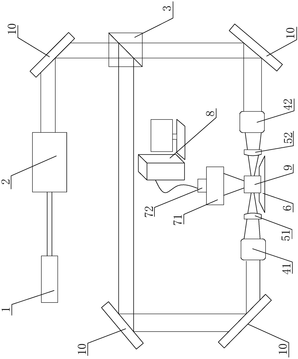
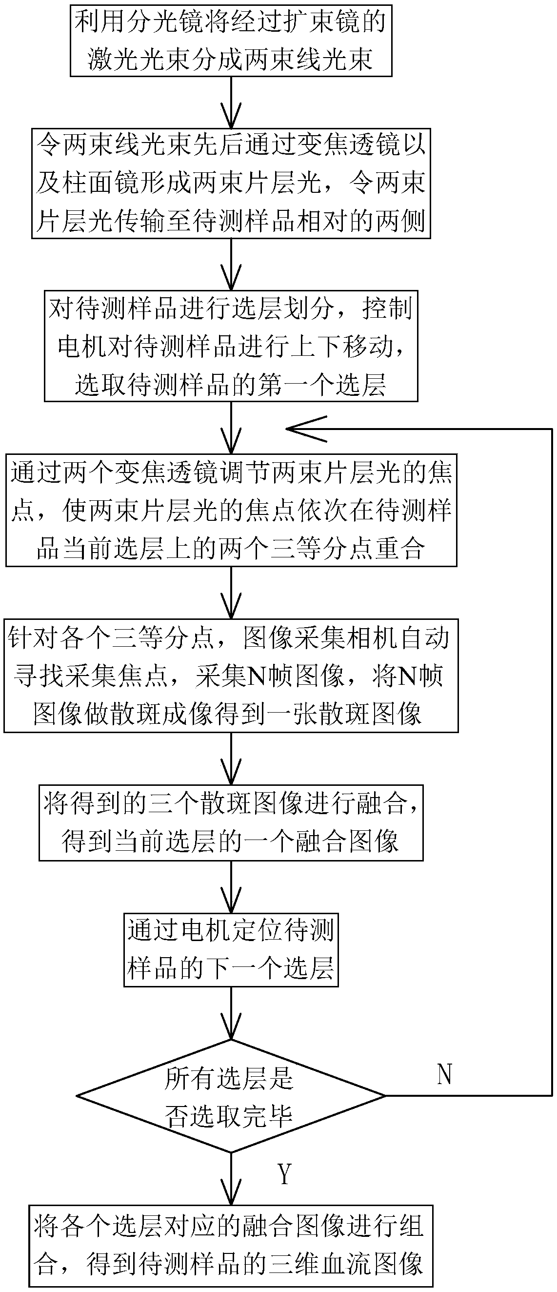
The invention discloses a selected-layer blood flow speckle imaging device and method based on sliced-layer light. The device comprises a laser light source, a beam expander, a beam splitter, a firstvarifocus lens, a second varifocus lens, a first cylindrical mirror, a second cylindrical mirror, a motor, an autofocus lens, an image acquisition camera and a processor; a laser beam output by the laser light source forms a first linear beam and a second linear beam through the beam expander and the beam splitter, the first linear beam sequentially passes through the first varifocus lens and thefirst cylindrical mirror and forms the first sliced-layer light, the second linear beam sequentially passes through the second varifocus lens and the second cylindrical mirror and forms the second sliced-layer light, and the first sliced-layer light and the second sliced-layer light enter the image acquisition camera through the autofocus lens after being absorbed by a sample to be tested. By means of the device, the two beams of sliced-layer light coincide at the trisection points of the sample to be tested to collect information of different selected layers of the sample to be tested; by means of the device, the complex fluorescent dyeing and detection operation is avoided, and the imaging operation is simple.

Description

technical field [0001] The present invention relates to the technical field of blood flow speckle imaging, in particular to a layer-selected blood flow speckle imaging device and method based on slice light. Background technique [0002] In terms of blood flow speckle imaging, there is currently an RFLSI Pro laser speckle blood flow imager from Beijing Giandel Technology Co., Ltd. that can monitor two-dimensional blood flow images at the micron level in real time, which can be measured and displayed intuitively. The moorFLPI-2 zoom laser speckle blood flow imaging system for tissue blood flow distribution, and the moorFLPI panoramic blood flow perfusion imaging system that can provide video rate up to 25 frames per second of blood flow changes in microvessels. [0003] In terms of slice light application imaging, it is now possible to detect senescent cells without complicated fluorescent labeling operations and detection processes. In the patent document with the publicati...

Claims


Application Information

Patent Timeline
IPC IPC(8): A61B5/026
CPCA61B5/0261
Inventor 张梦娇王茗祎曾亚光韩定安罗佳雄罗曦翁嘉瞬邓邦肯吴淋昌
Owner FOSHAN UNIVERSITY
Learn More