
Model for detecting malignant degrees of meningioma and application of model

A model, tumor technology, applied in the field of gene diagnosis and biology, can solve the problem of inability to diagnose tumors

Pending Publication Date: 2018-12-04
LISEN IMPRINTING DIAGNOSTICS WUXI CO LTD
View PDF8 Cites 1 Cited by
  • Summary
  • Abstract
  • Description
  • Claims
  • Application Information

AI Technical Summary

Problems solved by technology

[0006] At present, clinical research on imprinted genes is mainly based on methylation detection. The detection techniques mainly include restriction enzyme digestion, enrichment and sulfite conversion. There is a large variation in the effect of methylation, and even if an accurate methylation detection method exists, it cannot accurately diagnose the tumor

Method used


Image

  • Model for detecting malignant degrees of meningioma and application of model
  • Model for detecting malignant degrees of meningioma and application of model
  • Model for detecting malignant degrees of meningioma and application of model

Examples


Embodiment 1

[0150] Example 1 Imprinted gene analysis of thyroid cancer

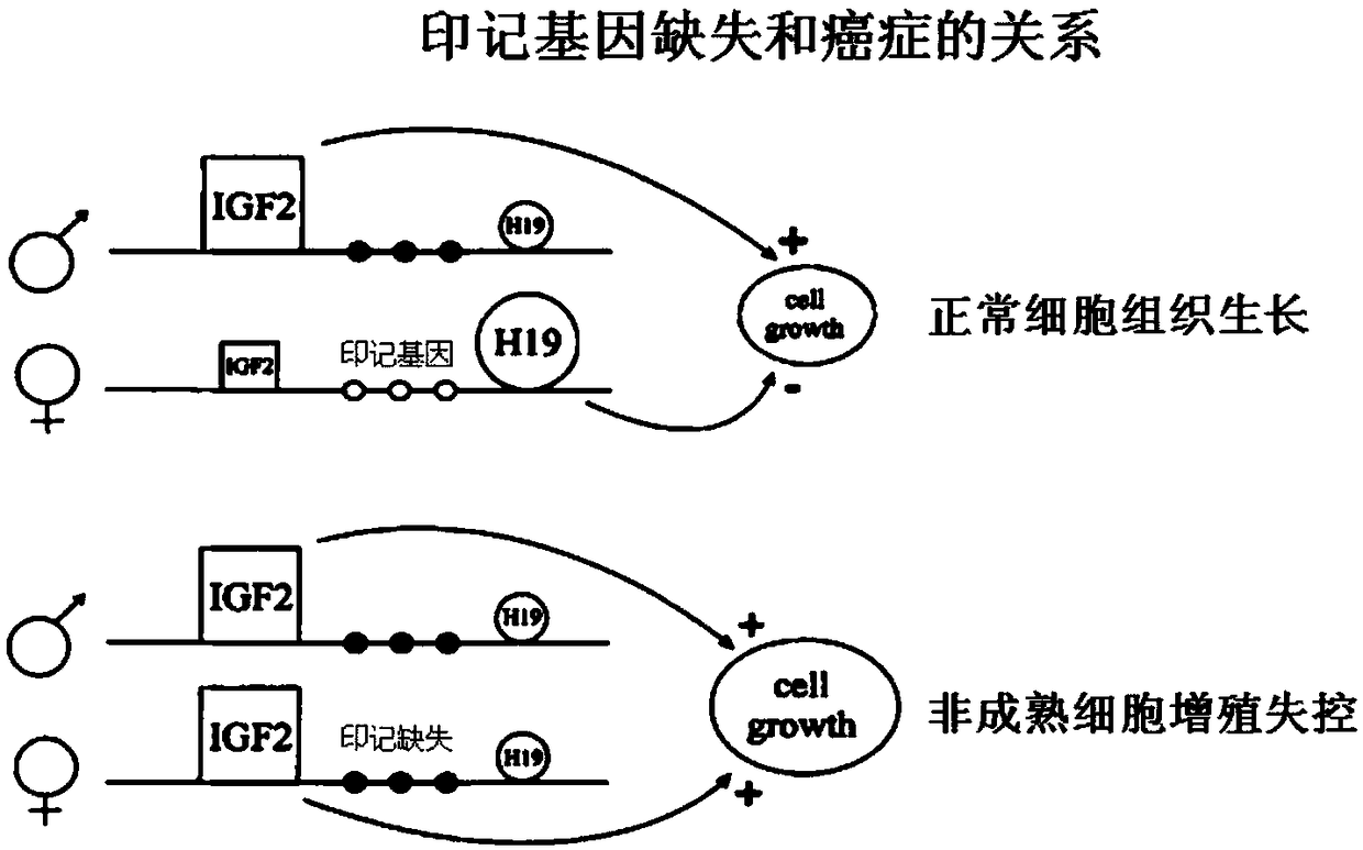
[0151] This embodiment provides a method for detecting thyroid cancer using imprinted genes, the relationship is as follows figure 1 Shown, described detection method comprises the steps:

[0152] (1) Obtain a tissue cell section (10 microns) of thyroid cancer, put it into the fixative solution described in patent ZL200710024048.6 and fix it to prevent RNA degradation. The fixation time is 24 hours, and it is embedded in paraffin (FFPE). Slides need to be detached with positive charges, and the slices are baked in a 40°C oven for more than 3 hours;

[0153] (2) Carry out dewaxing treatment according to the sample processing method of RNASCope to block the endogenous peroxidase activity in the sample, enhance permeability and expose RNA molecules;

[0154] (3) Design probes: design specific primers according to the imprinted gene sequence;

[0155] The design probes are based on imprinted genes Z1 (Gnas), Z2 (Igf2)...

Embodiment 2Z1-Z16

[0165] Example 2 Response sensitivity of Z1-Z16 imprinted genes to thyroid cancer

[0166] Obtain the tissue (10 microns) of 17 routine thyroid tumor patients, other detection methods are with embodiment 1, detection result is as follows Figure 4(a)-Figure 4(m) shown.

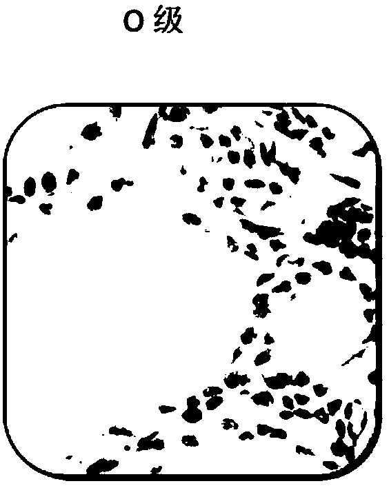
[0167] From Figure 4(a)-Figure 4(m) It can be seen that the proportion of imprint deletion and copy number abnormality of each probe in 17 thyroid tumor tissue samples presents a distribution from low to high. According to the distribution trend of different probes, we calculated the grade shown by the dotted line in the figure Standard, the imprint deletion and copy number abnormality of each probe can be divided into 5 grades from low to high, and the specific grades are as follows:

[0168] It can be seen from Figure 4(a) that for the imprinted gene Z1, the imprinted gene deletion expression is less than 15% and / or the imprinted gene copy number abnormal expression is less than 1.5% is grade 0, and the i...

Embodiment 3

[0187] Imprinted gene analysis of embodiment 3 skin cancer

[0188] Tissue samples of normal moles and skin malignant melanoma were obtained, fixed in 10% neutral formalin for more than 24 hours, other detection methods were the same as in Example 1, and the detection results were as follows Figure 5(a)-Figure 5(b) shown.

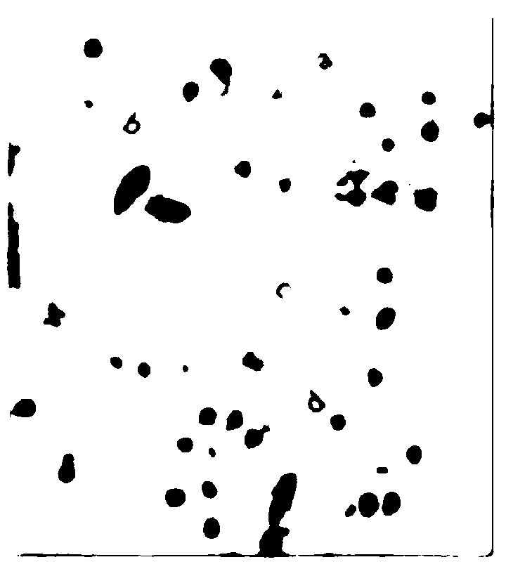
[0189] from Figure 5(a)-Figure 5(b) It can be seen that Figure 5(a) is a benign mole, and Figure 5(b) is a skin malignant melanoma. There are only individual cells with missing imprints in the mole, and no cells with abnormal copy number are found. There are a large number of skin malignant melanomas Cells with imprint deletions and copy number abnormalities.


PUM


Abstract

The invention relates to the field of genetic diagnosis, and in particular to a model for detecting malignant degrees of meningioma and an application of the model. The model, by calculating an imprinted gene deletion expression amount and an imprinted gene copy number abnormal expression amount, can grade changes of imprinted genes in meningioma. According to the detection model and the device provided by the invention, expression of imprint deletion on a sample of patients with meningioma is expressed by an intuitive method for the first time, and by virtue of an imprinted gene in-situ labeling method, changes of the imprinted genes can be objectively, intuitively and precisely detected out in early period; in addition, a quantization model can be provided; and therefore, the invention makes a huge contribution to the diagnosis of molecular pathology.

Description

technical field [0001] The present application relates to the field of biotechnology, to the field of gene diagnosis, and in particular to a model for detecting the degree of benign and malignant tumors and its application. Background technique [0002] According to the International Cancer Statistics, more than 8 million people die of cancer every year in the world. Therefore, early detection, diagnosis and treatment are particularly important. Cancer arises from complex genetic mutations and epigenetic changes that accumulate over time at the cellular level, ultimately leading to uncontrolled cell division. [0003] The diagnosis of benign and malignant cells by traditional pathology is based on the relationship between cell size, shape, invasion and surrounding cell tissue. It has great limitations in the discovery of early changes in cells (cancer), so the method of cancer diagnosis at the cell molecular level has once become a research hotspot. With the continuous in...

Claims


Application Information

Patent Timeline
IPC IPC(8): C12M1/34C12M1/00C12Q1/6841G06F19/20
CPCC12Q1/6841C12Q2563/107G16B25/10G16B40/20G16H50/20G16B5/00G06T7/0012G06T2207/10056G06T2207/30096C12Q1/6886
Inventor 成彤周宁
Owner LISEN IMPRINTING DIAGNOSTICS WUXI CO LTD
Learn More