Distinguishing Method of Fluorescence Microscopy Image Result

A technology of fluorescence microscope and discriminant method, which is applied in the field of fluorescence microscope images, can solve the problems of low work efficiency, high labor intensity of doctors, low accuracy of image recognition and judgment, etc.

Active Publication Date: 2021-06-08
AUTOBIO LABTEC INSTR CO LTD
View PDF4 Cites 0 Cited by
  • Summary
  • Abstract
  • Description
  • Claims
  • Application Information

AI Technical Summary

Problems solved by technology

At present, the recognition of fluorescent images is mostly done by doctors through naked eye observation to determine whether the samples are negative or positive. This not only has human-made visual errors, doctors' work experience and other factors, resulting in low accuracy of image recognition and judgment, but also the labor of doctors. High strength, low work efficiency

Method used


Image

  • Distinguishing Method of Fluorescence Microscopy Image Result
  • Distinguishing Method of Fluorescence Microscopy Image Result
  • Distinguishing Method of Fluorescence Microscopy Image Result

Examples


Embodiment Construction

[0019] The embodiments of the present invention will be described in detail below in conjunction with the accompanying drawings. This embodiment is implemented on the premise of the technical solution of the present invention, and detailed implementation methods and specific operating procedures are provided, but the scope of protection of the present invention is not limited to the following Described embodiment.

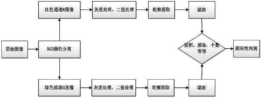
[0020] The fluorescence microscope image result discrimination method described in the present invention, such as figure 1 shown, follow the steps below:

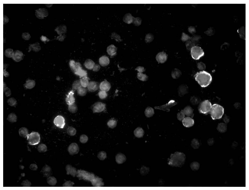
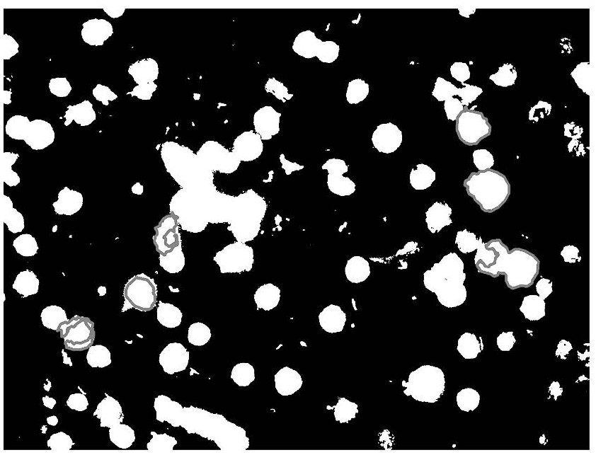
[0021] In the first step, the image of the sample taken by the fluorescence microscope (such as figure 2 shown) to separate the three channels of red, green and blue to obtain the red channel image (such as image 3 shown), the green channel image (such as Figure 4 As shown), the blue channel image; the blue channel image is discarded, and the red channel image and the green channel image are retained; the ...


PUM


Abstract

The invention discloses a method for discriminating the results of fluorescence microscope images. First, color-separate the red, green and blue channels of a sample image taken by a fluorescence microscope to obtain a red channel image, a green channel image, and a blue channel image; Perform grayscale processing and binary processing on the red and green channel images to obtain black and white binary images R1 and G1; 3. Extract the contours of the binary images R1 and G1 respectively to obtain multiple irregular polygonal images. Multiple irregular polygonal images are filtered; 4. When the number of cell images presented on the filtered binary image G1 is ≥ 1, and the presented cell images are similar to the cell images presented on the filtered binary image R1 When overlapping, it is judged that the sample image is positive; otherwise, it is judged that the sample image is negative. The invention has the advantages of realizing program batching and intelligently distinguishing negative and positive of tested samples, which greatly improves the working efficiency of doctors.

Description

technical field [0001] The invention relates to fluorescence microscope images, in particular to a method for judging the results of fluorescence microscope images. Background technique [0002] During the detection of pathogen markers, the sample is diluted and centrifuged, then placed in the detection instrument, and the processing steps such as slide loading, incubation, washing, and drying are automatically completed, and finally the slide is placed under a fluorescence microscope to take images , there are multiple holes (usually ten holes) on a glass slide, about ten fluorescence images need to be taken for each hole, and through image recognition of the ten fluorescence images of each hole, it is judged that the sample on each image is In the end, the judgment results of the ten images are summed up to obtain the negative and positive values ​​of the samples in the hole. At present, the recognition of fluorescent images is mostly done by doctors through naked eye obs...

Claims


Application Information

Patent Timeline
Patent Type & Authority Patents(China)
IPC IPC(8): G06T7/00G06T7/90G02B21/36
CPCG02B21/365G06T7/0012G06T2207/30024G06T7/90
Inventor 王超侯剑平赵万里刘聪
Owner AUTOBIO LABTEC INSTR CO LTD
Why Eureka
Try Eureka