
Thin layer magnetic resonance image reconstruction method based on deep learning

A magnetic resonance image and deep learning technology, which is applied in the generation of 2D images, image data processing, instruments, etc., can solve the problem that magnetic resonance images are difficult to achieve voxel-to-voxel registration, etc., and achieve good structure and details. Effect

Active Publication Date: 2018-10-09
FUDAN UNIV
View PDF3 Cites 27 Cited by
  • Summary
  • Abstract
  • Description
  • Claims
  • Application Information

AI Technical Summary

Problems solved by technology

Second, while MR images can be aligned to a normalized space, paired MR images are difficult to achieve voxel-to-voxel registration
Therefore, the reconstruction of thin-section MRI images will include complex processes of image registration and image reconstruction

Method used


Image

  • Thin layer magnetic resonance image reconstruction method based on deep learning
  • Thin layer magnetic resonance image reconstruction method based on deep learning
  • Thin layer magnetic resonance image reconstruction method based on deep learning

Examples


Embodiment 1

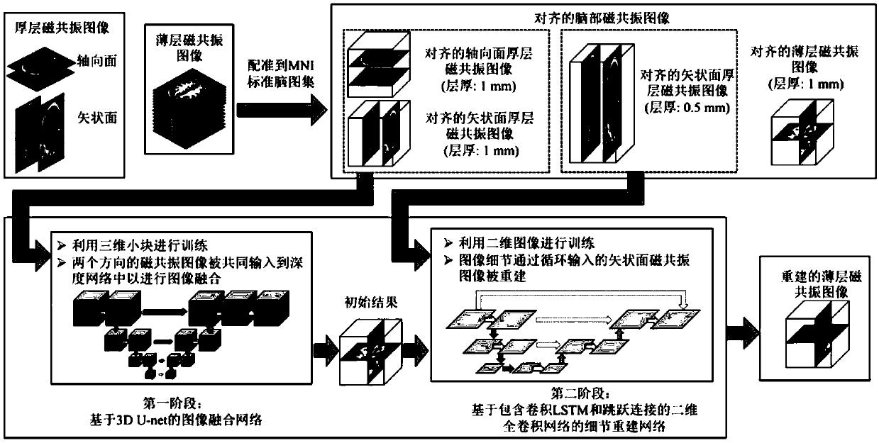
[0042] In the embodiment, a method for reconstructing thin-slice magnetic resonance images based on deep learning is proposed, which is represented by DeepVolume, such as figure 1 As shown, the specific steps are as follows.

[0043] 1) Acquisition of thick-slice magnetic resonance images in the axial and sagittal planes of the brain;

[0044] When collecting thick-slice magnetic resonance images, the pulse sequence of thick-slice axial plane magnetic resonance images is T1flair, the imaging plane is the axial plane, the sum of slice thickness and interslice distance is 6.5 mm, the number of slices is 19, and the pixel width is 0.47×0.47 mm, the repetition time is 2291ms, the echo time is 25mm, and the reversal time is 750ms. Thick-slice sagittal plane magnetic resonance image pulse sequence is T1flair, the imaging plane is the axial plane, the sum of slice thickness and slice distance is 6.5mm, the number of slices is 19, the pixel width is 0.47×0.47mm, and the repetition ti...


PUM


Abstract

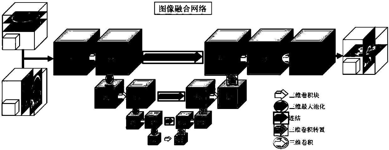
The invention discloses a thin layer magnetic resonance image reconstruction method based on deep learning. The method comprises the specific steps that 1) thick layer magnetic resonance images are collected on the axial and sagittal planes of a brain; 2) the thick layer magnetic resonance images are registered and normalized; 3) the paired and registered thick layer magnetic resonance images areused to train an image fusion network based on 3D U-net to generate a preliminary reconstruction result of a thin layer magnetic resonance image; and 4) the preliminary reconstruction result of the thin layer magnetic resonance image and a corresponding sagittal thick layer magnetic resonance image are used to train details to reconstruct the network and acquire a final reconstruction result. In the data set of magnetic resonance images of adolescent brains, the method provided by the invention can provide a better thin layer magnetic resonance image reconstruction result, and the reconstructed magnetic resonance image can better show the structure and detail of a brain. The estimation accuracy of gray matters, white matters and total brain volume in the magnetic resonance image can be greatly improved.

Description

technical field [0001] The invention belongs to the technical field of computer medical image processing, and in particular relates to a method for reconstructing thin-slice magnetic resonance images based on deep learning. Background technique [0002] Medical imaging technologies such as magnetic resonance imaging and computerized tomography (Computed Tomography, CT) can produce tomographic images of human body cross sections. The resulting imaging signal reconstructs a 3D volumetric image of the body part. The interval between slices is a key imaging parameter of magnetic resonance imaging. For example, thick-slice magnetic resonance images, also known as conventional magnetic resonance images, usually use a slice thickness of 5 mm to 8 mm. Thick-slice magnetic resonance images are often used for disease screening and initial diagnosis, but cannot be used to accurately describe brain structure and brain function, or for surgical planning and guidance, because many clinic...

Claims


Application Information

Patent Timeline
Patent Type & Authority Applications(China)
IPC IPC(8): G06T11/00G06N3/04
CPCG06T11/003G06N3/045
Inventor 汪源源余锦华李泽榉
Owner FUDAN UNIV
Learn More