Nanofiber membrane for wound healing and preparation method and application thereof
A composite nanofiber, wound healing technology, applied in bandages, absorbent pads, medical science and other directions, can solve problems such as being unsuitable for soft tissue wound healing, achieve good surface physical and chemical properties, reduce inflammatory response, and accelerate wound healing.
- Summary
- Abstract
- Description
- Claims
- Application Information
AI Technical Summary
Problems solved by technology
Method used
Image


Examples
Embodiment 1
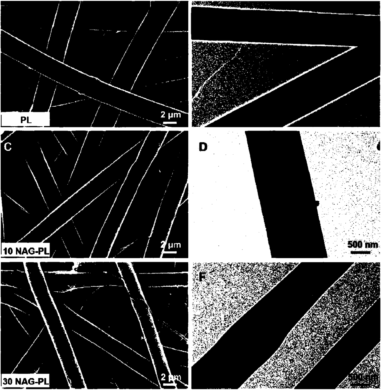
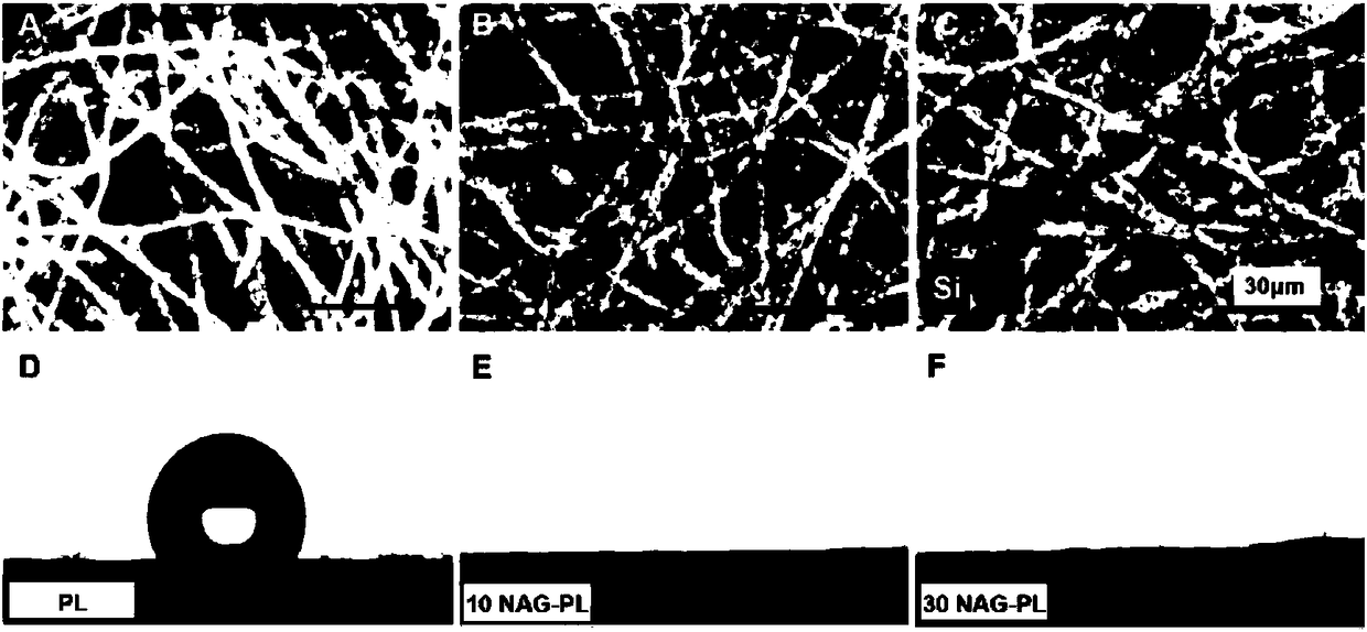
[0058] Example 1: Preparation of the composite nanofibrous membrane of the present invention for soft tissue wound healing
[0059] Dissolve PCL, gelatin and NAGEL in an appropriate amount of hexafluoroisopropanol (10%-25%, m / v) solvent at the same time, the addition amount of the three components is shown in each group in Table 1, and the magnetic stirring speed is 600rpm / min to obtain a uniform mixed electrospinning solution, and set up a control group A0 at the same time, see Table 1.
[0060] Table 1
[0061] test group
Polycaprolactone, wt%
Gelatin, wt%
NAGEL, wt%
Group A0 (PL)
50%
50%
0
Group A1 (30NAG-PL)
50%
50%
30%
Group A2 (10NAG-PL)
50%
50%
10%
[0062] Wherein, the PL means not containing NAGEL;
[0063] Wherein, the 10NAG-PL means that the content of NAGEL is 10%.
[0064] Wherein, said 30NAG-PL means that the content of NAGEL is 30%.
[0065] The prepared mixed electrospinning solution...
Embodiment 2
[0071] Example 2: Studying the Effect of the Composite Nanofiber Membrane for Soft Tissue Wound Healing on Cell Proliferation During Wound Repair
[0072] Each group of materials prepared in Example 1 was cut into a circular sheet with Φ=10 mm, soaked in 75% alcohol for 30 minutes, washed twice with sterilized PBS, and put into a 48-well plate for later use. Human umbilical vein endothelial cells, human fibroblasts, and human keratinocytes were planted on the sample surface, 6×10 per well 3 The culture medium used was ECM medium with 5% FBS and growth factors, DMEM medium with 10% FBS and 1640 medium with 10% FBS, the culture temperature was 37°C, and the culture atmosphere was 5% CO 2 air, and the medium was changed every two days. The CCK8 method was used to test the proliferation of cells on the surface of the material. After the cells were cultured for 1, 3, and 7 days, CCK8 solution was added to the 48-well plate in the dark, and cultured at 37° C. for another 2-4 hour...
Embodiment 3
[0076] Example 3: Studying the Effect of the Composite Nanofibrous Membrane for Soft Tissue Wound Healing on the Migration and Adhesion of Human Angiogenesis Cells
[0077] Digest the HUVEC cells in the logarithmic phase with trypsin. After digestion, inoculate the cells in a 24-well plate. When the cell density is about 90% at the bottom of the culture dish, gently draw a straight line with a sterile medium pipette tip. , aspirate the culture medium, and wash twice with PBS. A transwell chamber is added to the upper part of the 24-well plate, and the material prepared in Example 1 is added therein, and the culture medium is filled up and the cells are placed in a 5% CO2 incubator for culture. After culturing for 6-8 hours, take pictures under the OLYMPUS microscope, and count the number of migrated cells.
[0078] Add different materials to the bottom of a 24-well cell culture plate equipped with Boyden chambers. Add 600 μL of endothelial cell culture medium containing 10% s...
PUM
 Abstract
Description
Claims
Application Information
 - R&D
- Intellectual Property
- Life Sciences
- Materials
- Tech Scout
- Unparalleled Data Quality
- Higher Quality Content
- 60% Fewer Hallucinations
Browse by: Latest US Patents, China's latest patents, Technical Efficacy Thesaurus, Application Domain, Technology Topic, Popular Technical Reports.
© 2025 PatSnap. All rights reserved.Legal|Privacy policy|Modern Slavery Act Transparency Statement|Sitemap|About US| Contact US: help@patsnap.com