Nerve conduit capable of improving axonal regeneration orderliness and preparation method thereof
A nerve conduit and axon regeneration technology, applied in the field of nerve conduits, to achieve the effect of improving orderliness
- Summary
- Abstract
- Description
- Claims
- Application Information
AI Technical Summary
Problems solved by technology
Method used
Image
Examples
Embodiment 1
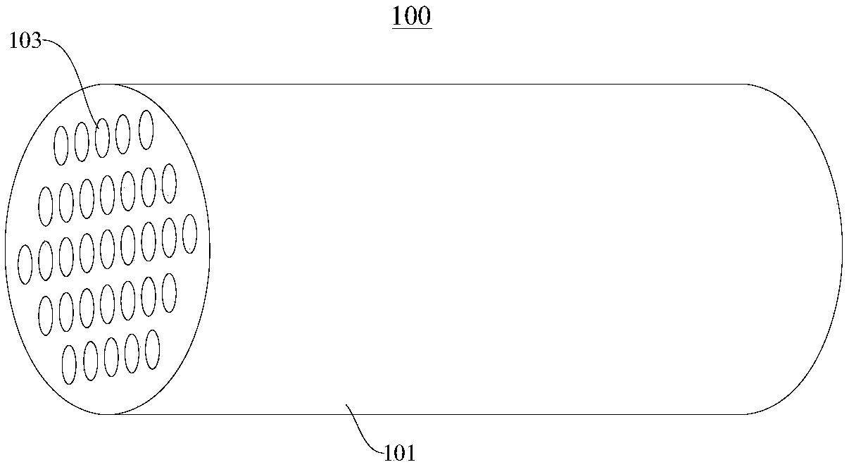
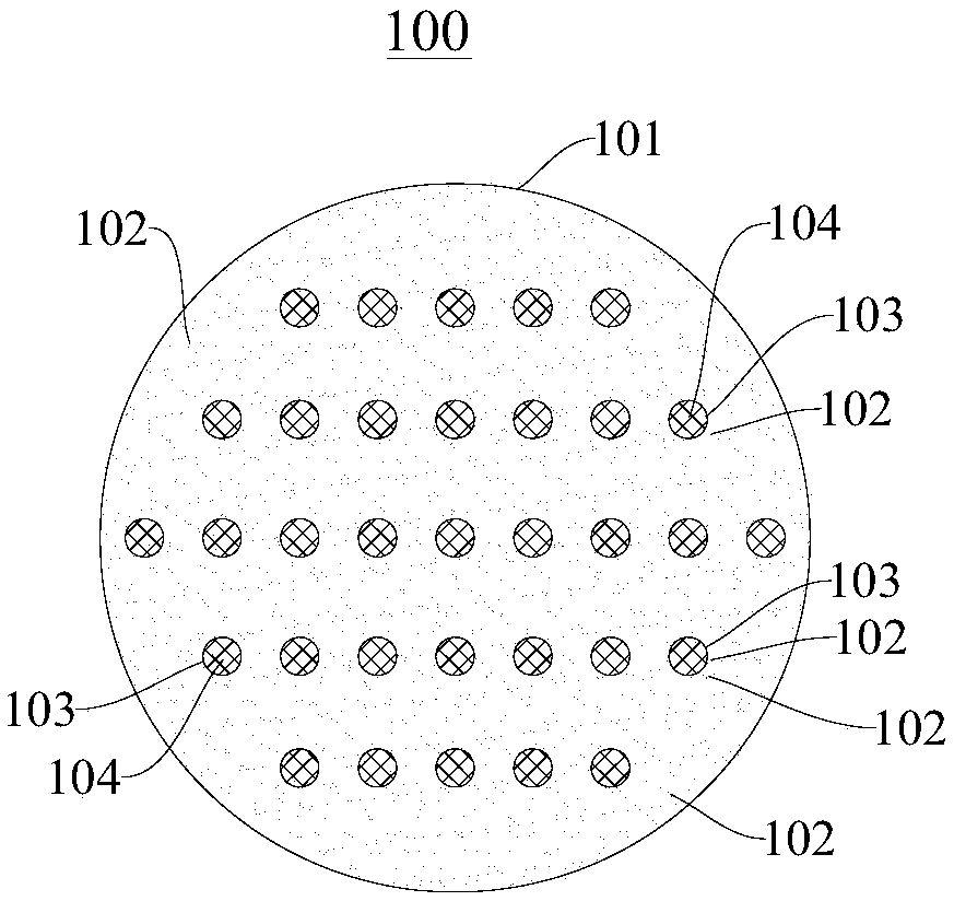
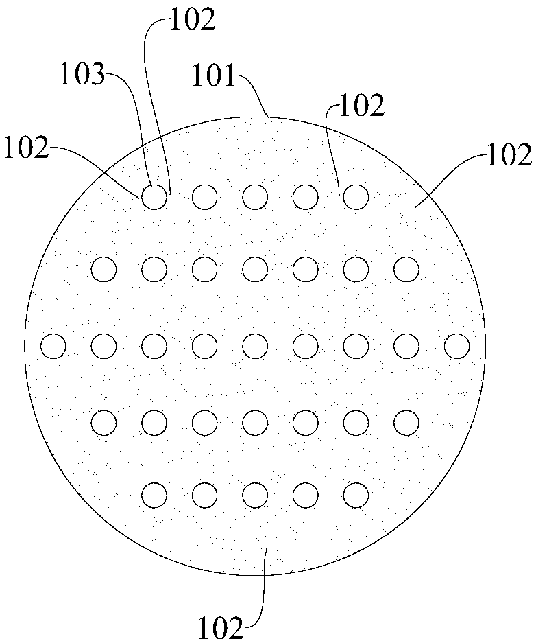
[0043] Please refer to Figure 1-Figure 4 , the nerve guide 100 that can improve the orderliness of axon regeneration provided by this embodiment includes:
[0044]The tube body 101 made of gelatin and the axon-inhibiting molecules distributed in the tube body 101 that have an inhibitory effect on axon regeneration are chondroitin sulfate proteoglycans 102 (CSPGs) in this embodiment.
[0045] Wherein, the inner wall of the tubular body 101 has a plurality of nerve regeneration channels 103 extending along the length direction of the tubular body 101; The channels 103 are distributed in the inside of the tube body 101 in a manner of distributing along the length direction.
[0046] In addition, the nerve regeneration channel 103 is filled with hydrogel 104 that supports and promotes the growth of nerve fibers.
[0047] In addition, the outer diameter of the nerve guide 100 is about 2-5 mm; the diameter of the nerve regeneration channel 103 is about 100-300 μm. Of course, in ...
experiment example 1
[0060] Effect of CSPGs distribution on axon regeneration
[0061] Methods: (Adult SD rats were modeled with sciatic nerve clamp injury, and the SD rats were euthanized 7 days after the injury, and the injured sciatic nerve was taken, fixed in paraformaldehyde, and frozen sectioned, and the longitudinal field of view was taken, followed by immunofluorescence The staining method specifically marked axons (NF200) and chondroitin sulfate proteoglycans (CSPGs), and the results were as follows Figure 5 shown.
[0062] from Figure 5 It can be seen that the effect of axon regeneration after 7 days of injury repair is related to the distribution of CSPGs. Where the distribution of CSPGs is regular, the axon runs smoothly (arrow), while where the distribution of CSPGs is chaotic, the regeneration of axon is disordered (as indicated by the arrow in the figure).
experiment example 2
[0064] Flat CSPGs interface guides axonal steering
[0065] Method: Smear CSPGs on the bottom of the culture dish pretreated with poly-lysine to form the boundary of CSPGs, then plant DRG neurons in the culture dish, fix the cells with paraformaldehyde after a period of culture, and perform immunofluorescence staining Mark the axons, observe the growth of the axons at the boundary of CSPGs under a microscope, the results are as follows Image 6 shown.
[0066] Image 6 -B1 shows that DRG neurons grow and form distinct axonal boundaries in culture plates coated with CSPGs ( Image 6 -B1 indicated by the arrow); Image 6 -C shows that NF200 immunofluorescence staining shows that axons turn around near the CSPGs interface, and the enlarged images of rectangular boxes (corresponding to 1, 2, 3, 4) show the angle of axon turning; Image 6 -D shows that the proportion of axons that are turned (0.88±0.09) by the CSPGs interface is much higher than the proportion of axons that are...
PUM
| Property | Measurement | Unit |
|---|---|---|
| Diameter | aaaaa | aaaaa |
| Diameter | aaaaa | aaaaa |
| Outer diameter | aaaaa | aaaaa |
Abstract
Description
Claims
Application Information
 - R&D Engineer
- R&D Manager
- IP Professional
- Industry Leading Data Capabilities
- Powerful AI technology
- Patent DNA Extraction
Browse by: Latest US Patents, China's latest patents, Technical Efficacy Thesaurus, Application Domain, Technology Topic, Popular Technical Reports.
© 2024 PatSnap. All rights reserved.Legal|Privacy policy|Modern Slavery Act Transparency Statement|Sitemap|About US| Contact US: help@patsnap.com


