Eureka AIR delivers breakthrough ideas for toughest innovation challenges, trusted by R&D personnel around the world.

Method for establishing in-vitro model capable of simulating barrier functions of pulmonary epithelial cells and capillary endothelial cells during acute lung injury

An endothelial cell barrier, lung epithelial cell technology, applied in vascular endothelial cells, epidermal cells/skin cells, artificial cell constructs, etc., can solve problems that do not involve barrier function, etc.

Inactive Publication Date: 2018-05-04
KUNMING MEDICAL UNIVERSITY
View PDF0 Cites 7 Cited by
  • Summary
  • Abstract
  • Description
  • Claims
  • Application Information

AI Technical Summary

Problems solved by technology

Zhou Changxi et al. used the Transwell co-culture system to create a co-culture model of human type II alveolar epithelial cell line A549 and human pulmonary microvascular endothelial cell line HPMECs, and used it to study the effect of flagellin stimulation on the expression of TNF-α in lung epithelial cells. impact, but Zhou Changxi’s research did not involve the barrier function of lung epithelial / endothelial cells in the most common endotoxin ALI in clinical practice. Does infection induce inflammation in pulmonary vascular endothelial cells and transmit cascades to alveolar epithelial cells

Method used


Image

  • Method for establishing in-vitro model capable of simulating barrier functions of pulmonary epithelial cells and capillary endothelial cells during acute lung injury
  • Method for establishing in-vitro model capable of simulating barrier functions of pulmonary epithelial cells and capillary endothelial cells during acute lung injury
  • Method for establishing in-vitro model capable of simulating barrier functions of pulmonary epithelial cells and capillary endothelial cells during acute lung injury

Examples


Embodiment 1

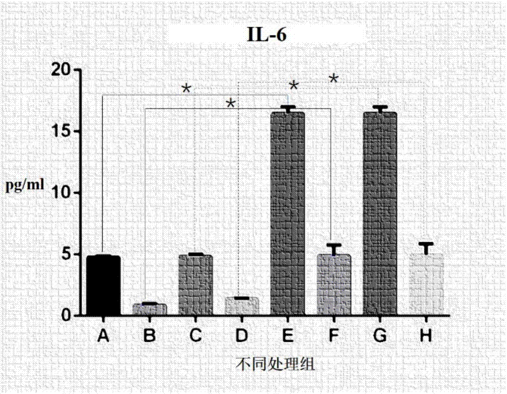
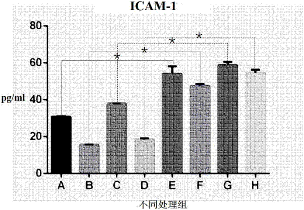
[0021] In this example, a co-culture model of mouse TC-1 lung epithelial cells and mouse C3H / 10T1 / 2 microvascular endothelial cells was made in the Transwell co-culture system, and the lung epithelial cells / microvascular endothelial cells were co-cultured by adjusting the cell culture environment. The culture model can express inflammatory cytokines (IL6) and inflammatory cell infiltration-associated protein (ICAM-1) under endotoxin stimulation.

[0022] The process of making a lung epithelial cell / microvascular endothelial cell co-culture model is as follows:

[0023] Using the Transwell co-culture system, the mouse C3H / 10T1 / 2 microvascular endothelial cell line infected with 1ug / mL LPS was mixed with 4 × 10 4 / cm 2 The concentration of 1.5mL was inoculated on the lower layer of the Transwell chamber, and cultured for 5h. The mouse-derived TC-1 lung epithelial cell line was mixed with 2×10 4 / cm 2 Inoculate 1mL on the upper layer of the small chamber. The lower cell cult...

Embodiment 2

[0031] Example 2 Changing the concentration of LPS

[0032] After pre-experimental exploration, it was found that the cells in the co-culture system induced by the final concentration of 0.8-1.2 μg / mL LPS could survive and grow well. Concentrations below 0.8 μg / mL will result in failure to cause damage or a large gap between the degree of damage and the situation of acute lung injury in vivo. Above 1.2μg / mL, it will lead to cell death.

Embodiment 3

[0033] Example 3 Changing the lower cell culture medium

[0034] The established lower medium is the most suitable culture environment for the growth of endothelial cells. Changing the components in the medium, or missing a certain component, or changing the amount and ratio of components will lead to low survival rate of endothelial cells, slow proliferation, or even death. The cell density required for the co-culture of the two types of cells could not be reached, and the cell culture results are shown in Table 1.

[0035] Table 1

[0036]

[0037] Typically, cells die the next day when no FBS is added. In the absence of 90-95U / mL heparin, there is little effect on cell viability and density. Too high concentrations of penicillin sodium and streptomycin sulfate may cause cell death, and too low concentrations may cause bacterial contamination.


PUM

PropertyMeasurementUnit
concentrationaaaaaaaaaa

Abstract

The invention discloses a method for establishing an in-vitro model capable of simulating barrier functions of pulmonary epithelial cells and capillary endothelial cells during acute lung injury. Themethod comprises the following steps: (1) inoculating endotoxin-infected mouse C3H / 10T1 / 2 microvascular endothelial cell strains into lower culture of a cell by using a Transwell co-culture system; inoculating mouse-derived TC-1 pulmonary epithelial cells into the upper layer of the cell, co-culturing for a period of time, and collecting the cell culture supernatant; and (2) detecting conditions of barrier synthesis of pulmonary epithelial cells / capillary endothelial cells in the cell culture supernatant and expressions of inflammatory factor and inflammatory cell infiltration associated proteins, and establishing a pulmonary epithelial cell / capillary endothelial cell co-culture model applicable to endotoxic acute lung injury study. The model established by the method disclosed by the invention is close to a cell barrier function during in-vivo endotoxic acute lung injury, and can provide a reliable model basis for clinical acute lung injury study.

Description

technical field [0001] The invention relates to the technical field of cell model establishment, in particular to an in vitro model establishment method for simulating the barrier function of lung epithelial cells and capillary endothelial cells during acute lung injury. Background technique [0002] Endotoxin-induced acute lung injury (acute lung injury, ALI) is a common critical illness clinically. The pathogenesis of ALI is diverse, and the endotoxin lipopolysaccharide (LPS) accounts for the vast majority, which can be divided into LPS direct injury and LPS indirect injury (imbalance of pro-inflammatory / anti-inflammatory response, imbalance of pro-coagulant / anti-coagulant response , oxidative stress, apoptosis disorder, etc.), alveolar-capillary barrier III, leading to increased capillary permeability, neutrophil infiltration and uncontrolled release of inflammatory mediators, and aggregation at the injury site induces a series of symptom. ALI is the result of uncontrol...

Claims


Application Information

Patent Timeline
Patent Type & Authority Applications(China)
IPC IPC(8): C12N5/071
CPCC12N5/069C12N5/0625C12N2502/09C12N2502/28
Inventor 马微李力燕郭建辉杨金伟代云飞王先斌刘矿嫔张同王嘉蔚
Owner KUNMING MEDICAL UNIVERSITY
Why Eureka
Eureka Blog
Learn More