Evaluation method of paracancerous collagenous tissue of early gastric cancer excision specimen
A technology for early gastric cancer and paracancerous collagen, applied in the field of pathological identification, can solve problems such as reducing the quality of life of patients, achieve the effect of reducing the probability of tumor recurrence, broad clinical application prospects, and mature technology
- Summary
- Abstract
- Description
- Claims
- Application Information
AI Technical Summary
Problems solved by technology
Method used
Image
Examples
Embodiment 1
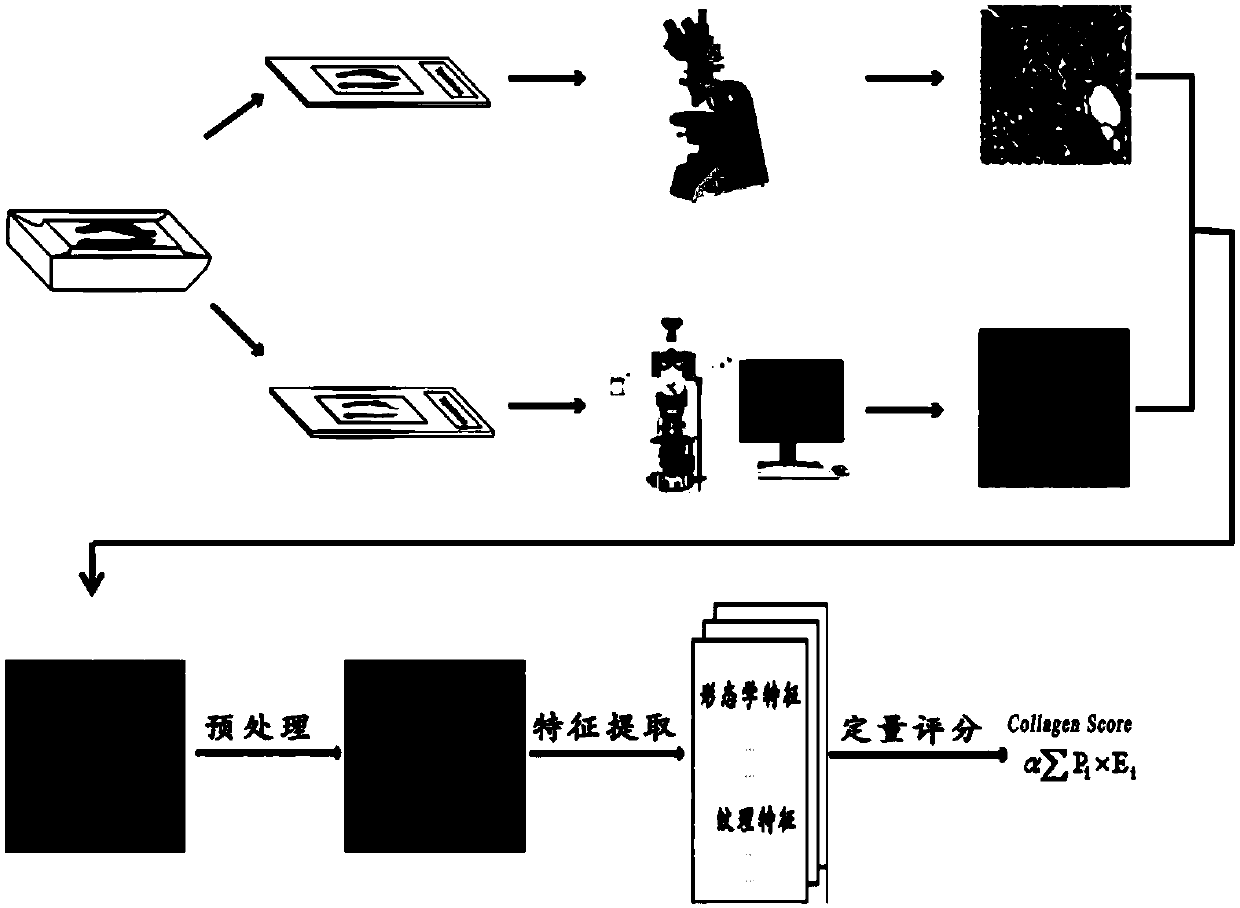
[0051] Such as figure 1 Shown, a kind of paracancerous collagen tissue evaluation method of early gastric cancer resection specimen, described method comprises the following steps:
[0052] (a) Sample preparation: 3 consecutive tissue sections with a thickness of 3 μm were cut from the paracancerous part of the early gastric cancer specimen for routine wax block preparation, one for hematoxylin-eosin staining, and the other for pathological Masson staining , and the remaining one was placed in a -86°C refrigerator for refrigerated storage and used as a multiphoton imaging slice to be tested;
[0053] (b) Marking the imaging area: Observe the area in the hematoxylin-eosin-stained section and the Masson-stained section that matches the collagen adjacent to the early gastric cancer under an ordinary optical microscope, and compare it with the multiphoton imaging section to be tested in step (a). For comparison, mark the corresponding region on the multiphoton imaging slice to be...
Embodiment 2
[0057] A method for evaluating paracancerous collagen tissue of a resection specimen of early gastric cancer, the method comprising the following steps:
[0058] (a) Sample preparation: 3 consecutive tissue sections with a thickness of 8 μm were cut from the paracancerous part of the early gastric cancer specimen for routine wax block preparation, one for hematoxylin-eosin staining, and the other for pathological Masson staining , and the remaining one was placed in a -86°C refrigerator for refrigerated storage and used as a multiphoton imaging slice to be tested;
[0059] (b) Marking the imaging area: Observe the area in the hematoxylin-eosin-stained section and the Masson-stained section that matches the collagen adjacent to the early gastric cancer under an ordinary optical microscope, and compare it with the multiphoton imaging section to be tested in step (a). For comparison, mark the corresponding region on the multiphoton imaging slice to be measured to obtain the marke...
Embodiment 3
[0063] A method for evaluating paracancerous collagen tissue of a resection specimen of early gastric cancer, the method comprising the following steps:
[0064] (a) Sample preparation: 3 consecutive tissue sections with a thickness of 5 μm were cut from the paracancerous part of the early gastric cancer specimen for routine wax block preparation, one was taken for hematoxylin-eosin staining, and the other was used for pathological Masson staining , and the remaining one was placed in a -86°C refrigerator for refrigerated storage and used as a multiphoton imaging slice to be tested;
[0065] (b) Marking the imaging area: Observe the area in the hematoxylin-eosin-stained section and the Masson-stained section that matches the collagen adjacent to the early gastric cancer under an ordinary optical microscope, and compare it with the multiphoton imaging section to be tested in step (a). For comparison, mark the corresponding region on the multiphoton imaging slice to be measured ...
PUM
| Property | Measurement | Unit |
|---|---|---|
| thickness | aaaaa | aaaaa |
Abstract
Description
Claims
Application Information
 - R&D
- Intellectual Property
- Life Sciences
- Materials
- Tech Scout
- Unparalleled Data Quality
- Higher Quality Content
- 60% Fewer Hallucinations
Browse by: Latest US Patents, China's latest patents, Technical Efficacy Thesaurus, Application Domain, Technology Topic, Popular Technical Reports.
© 2025 PatSnap. All rights reserved.Legal|Privacy policy|Modern Slavery Act Transparency Statement|Sitemap|About US| Contact US: help@patsnap.com


