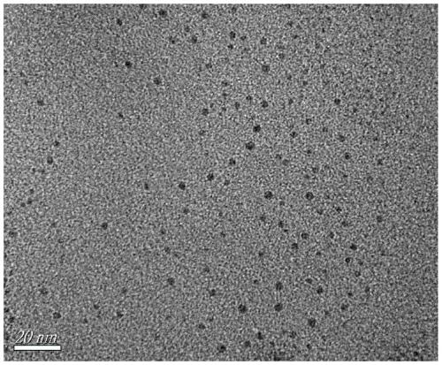
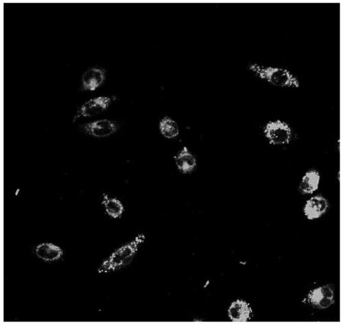
Methoxyfluoroboron pyrrole-nucleic acid-ferric oxide complex and preparation method
A technology of methoxyfluoroboron pyrrole and ferric tetroxide, which is applied in the field of fluorescent imaging agents
- Summary
- Abstract
- Description
- Claims
- Application Information
AI Technical Summary
Problems solved by technology
Method used
Image
Examples
Embodiment example 1
[0016] Implementation case 1 (best synthesis condition):
[0017] DNA 40-1 : 5'-GAGGAGACAACAACAGCGCGCGC-3' and DNA 40-1D : 5'-GCGCGCGCACAACAACAGAGGAG-3' small molecule nucleic acid as a template, the DNA 40-1 Mix with ferric chloride solution in sodium citrate buffer (pH=9, 10mM) to obtain No. 1 mixed solution, DNA 40-1 with Fe 3+ The concentrations were 6.4μM and 2000μM, and the DNA 40-1D , vitamin C solution and ferrous ammonium sulfate solution are mixed in sodium citrate buffer (pH=9, 10mM) to obtain No. 2 mixed solution, DNA 40-1D and, Vitamin C and Fe 2+The concentrations were 6.4μM, 1000μM and 1110μM respectively; then the above two mixed solutions were transferred to the water bath for heating, and the temperature rose to 67°C and stabilized for three minutes. Transfer 20 μL of ammonia water (pH=12) to a shaker for 30 minutes, then slowly cool down to room temperature for 5 hours; finally, drop 20 μL of vitamin C (10 mM) solution into the above solution, mix well...
Embodiment example 2
[0019] DNA 40-1 : 5'-GAGGAGACAACAACAGCGCGCGC-3' and DNA 40-1D : 5'-GCGCGCGCACAACAACAGAGGAG-3' small molecule nucleic acid as a template, the DNA 40-1 Mix with ferric chloride solution in sodium citrate buffer (pH=5, 10mM) to obtain No. 1 mixed solution, DNA 40-1 with Fe 3+ The concentrations were 6.4μM and 2000μM, and the DNA 40-1D , vitamin C solution and ammonium ferrous sulfate solution are mixed in sodium citrate buffer solution (pH=5, 10mM), obtain No. 2 mixed solution, DNA 40-1D and, Vitamin C and Fe 2+ The concentrations were 6.4 μM, 2000 μM and 2000 μM respectively; then the above two mixed solutions were transferred to a water bath for heating, and the temperature rose to 70°C and stabilized for three minutes, and the No. 1 and No. Transfer 20 μL of ammonia water (pH=12) to a shaker for 2 hours, then slowly cool down to room temperature for 6 hours; finally, drop 20 μL of vitamin C (10 mM) solution into the above solution, mix well and store in the dark for 5 min...
Embodiment example 3
[0021] DNA 40-1 : 5'-GAGGAGACAACAACAGCGCGCGC-3' and DNA 40-1D : 5'-GCGCGCGCACAACAACAGAGGAG-3' small molecule nucleic acid as a template, the DNA 40-1 Mixed with ferric chloride solution in sodium citrate buffer (pH=7, 10mM) to obtain No. 1 mixed solution, DNA 40-1 with Fe 3+ The concentrations were 6.4μM and 2000μM, and the DNA 40-1D , vitamin C solution and ammonium ferrous sulfate solution are mixed in sodium citrate buffer solution (pH=7, 10mM), obtain No. 2 mixed solution, DNA 40-1D and, Vitamin C and Fe 2+ The concentrations were 6.4 μM, 2000 μM and 2000 μM respectively; then the above two mixed solutions were transferred to a water bath for heating, and the temperature rose to 70°C and stabilized for three minutes, and the No. 1 and No. 2 mixed solutions were mixed evenly, and 20 μL Ammonia water (pH=12), transferred to a shaker for 2 hours, slowly cooled to room temperature, the cooling time was 6 hours; finally, 20 μL of vitamin C (10 mM) solution was added dropwi...
PUM
 Abstract
Description
Claims
Application Information
 - R&D
- Intellectual Property
- Life Sciences
- Materials
- Tech Scout
- Unparalleled Data Quality
- Higher Quality Content
- 60% Fewer Hallucinations
Browse by: Latest US Patents, China's latest patents, Technical Efficacy Thesaurus, Application Domain, Technology Topic, Popular Technical Reports.
© 2025 PatSnap. All rights reserved.Legal|Privacy policy|Modern Slavery Act Transparency Statement|Sitemap|About US| Contact US: help@patsnap.com


